Molecular recognition of trehalose and trehalose analogues by Mycobacterium tuberculosis LpqY-SugABC.
Liang, J., Liu, F., Xu, P., Shangguan, W., Hu, T., Wang, S., Yang, X., Xiong, Z., Yang, X., Guddat, L.W., Yu, B., Rao, Z., Zhang, B.(2023) Proc Natl Acad Sci U S A 120: e2307625120-e2307625120
- PubMed: 37603751 Search on PubMedSearch on PubMed Central
- DOI: https://doi.org/10.1073/pnas.2307625120
- Primary Citation Related Structures:
8JA7, 8JA8, 8JA9, 8JAA, 8JAB, 8JAC, 8JAD - PubMed Abstract:
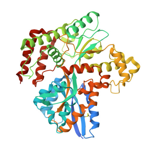
Trehalose plays a crucial role in the survival and virulence of the deadly human pathogen Mycobacterium tuberculosis ( Mtb ). The type I ATP-binding cassette (ABC) transporter LpqY-SugABC is the sole pathway for trehalose to enter Mtb . The substrate-binding protein, LpqY, which forms a stable complex with the translocator SugABC, recognizes and captures trehalose and its analogues in the periplasmic space, but the precise molecular mechanism for this process is still not well understood. This study reports a 3.02-Å cryoelectron microscopy structure of trehalose-bound Mtb LpqY-SugABC in the pretranslocation state, a crystal structure of Mtb LpqY in a closed form with trehalose bound and five crystal structures of Mtb LpqY in complex with different trehalose analogues. These structures, accompanied by substrate-stimulated ATPase activity data, reveal how LpqY recognizes and binds trehalose and its analogues, and highlight the flexibility in the substrate binding pocket of LpqY. These data provide critical insights into the design of trehalose analogues that could serve as potential molecular probe tools or as anti-TB drugs.
- State Key Laboratory of Medicinal Chemical Biology, Nankai University, Tianjin 300353, China.
Organizational Affiliation: