8J54

Crystal structure of RXR/DR2 complex


Experimental Data Snapshot

  • Method: X-RAY DIFFRACTION
  • Resolution: 2.72 Å
  • R-Value Free: 0.273 
  • R-Value Work: 0.217 
  • R-Value Observed: 0.223 

wwPDB Validation   3D Report Full Report


This is version 1.2 of the entry. See complete history


Literature

Structural characterization of the DNA binding mechanism of retinoic acid-related orphan receptor gamma.

Jiang, L.Liu, X.Liang, X.Dai, S.Wei, H.Guo, M.Chen, Z.Xiao, D.Chen, Y.

(2024) Structure 32: 467-475.e3

  • DOI: https://doi.org/10.1016/j.str.2024.01.004
  • Primary Citation of Related Structures:  
    7XVN, 8J54

  • PubMed Abstract: 

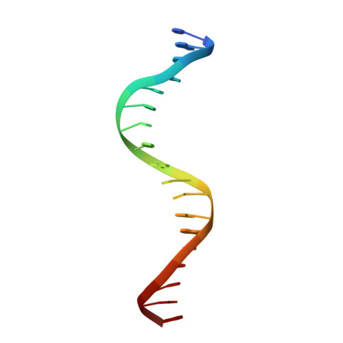
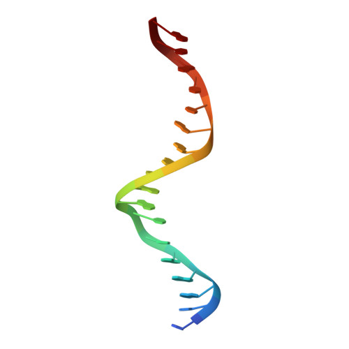
    Retinoic acid-related orphan receptor gamma (RORγ) plays critical roles in regulating various biological processes and has been linked to immunodeficiency disorders and cancers. DNA recognition is essential for RORγ to exert its functions. However, the underlying mechanism of the DNA binding by RORγ remains unclear. In this study, we present the crystal structure of the complex of RORγ1 DNA-binding domain (RORγ1-DBD)/direct repeat DNA element DR2 at 2.3 Å resolution. We demonstrate that RORγ1-DBD binds the DR2 motif as a homodimer, with the C-terminal extension (CTE) region of RORγ1-DBD contributing to the DNA recognition and the formation of dimeric interface. Further studies reveal that REV-ERB-DBD and RXR-DBD, also bind the DR2 site as a homodimer, while NR4A2-DBD binds DR2 as a monomer. Our research uncovers a binding mechanism of RORγ1 to the DR2 site and provides insights into the biological functions of RORγ1 and the broader RORs subfamily.


  • Organizational Affiliation

    Department of Pathology, NHC Key Laboratory of Cancer Proteomics, State Local Joint Engineering Laboratory for Anticancer Drugs, Department of Oncology, National Clinical Research Center for Geriatric Disorders, Xiangya Hospital, Central South University, Changsha, Hunan, China.


Macromolecules

Find similar proteins by:  (by identity cutoff)  |  3D Structure
Entity ID: 3
MoleculeChains Sequence LengthOrganismDetailsImage
Retinoic acid receptor RXR
C, D, E, F
83Mus musculusMutation(s): 0 
Gene Names: RxraRXR alpha
UniProt
Find proteins for P28700 (Mus musculus)
Explore P28700 
Go to UniProtKB:  P28700
Entity Groups  
Sequence Clusters30% Identity50% Identity70% Identity90% Identity95% Identity100% Identity
UniProt GroupP28700
Sequence Annotations
Expand
  • Reference Sequence

Find similar nucleic acids by:  Sequence   |   3D Structure  

Entity ID: 1
MoleculeChains LengthOrganismImage
DNA (5'-D(*CP*AP*TP*GP*AP*CP*CP*TP*AP*CP*TP*GP*AP*CP*CP*TP*AP*G)-3')A,
G [auth N]
18Homo sapiens
Sequence Annotations
Expand
  • Reference Sequence

Find similar nucleic acids by:  Sequence   |   3D Structure  

Entity ID: 2
MoleculeChains LengthOrganismImage
DNA (5'-D(*CP*TP*AP*GP*GP*TP*CP*AP*GP*TP*AP*GP*GP*TP*CP*AP*TP*G)-3')B,
H [auth P]
18Homo sapiens
Sequence Annotations
Expand
  • Reference Sequence
Experimental Data & Validation

Experimental Data

  • Method: X-RAY DIFFRACTION
  • Resolution: 2.72 Å
  • R-Value Free: 0.273 
  • R-Value Work: 0.217 
  • R-Value Observed: 0.223 
  • Space Group: P 21 21 21
Unit Cell:
Length ( Å )Angle ( ˚ )
a = 36.239α = 90
b = 63.646β = 90
c = 244.649γ = 90
Software Package:
Software NamePurpose
PHENIXrefinement
HKL-2000data reduction
autoPROCdata scaling
PHASERphasing

Structure Validation

View Full Validation Report


Entry History & Funding Information

Deposition Data


Funding OrganizationLocationGrant Number
National Natural Science Foundation of China (NSFC)China81974074 and 82172654

Revision History  (Full details and data files)

  • Version 1.0: 2024-01-17
    Type: Initial release
  • Version 1.1: 2024-02-14
    Changes: Database references
  • Version 1.2: 2024-04-17
    Changes: Database references