8IXB

GMPCPP-Alpha1A/Beta2A-microtubule decorated with kinesin seam region


Experimental Data Snapshot

  • Method: ELECTRON MICROSCOPY
  • Resolution: 4.20 Å
  • Aggregation State: FILAMENT 
  • Reconstruction Method: SINGLE PARTICLE 

wwPDB Validation   3D Report Full Report


This is version 1.2 of the entry. See complete history


Literature

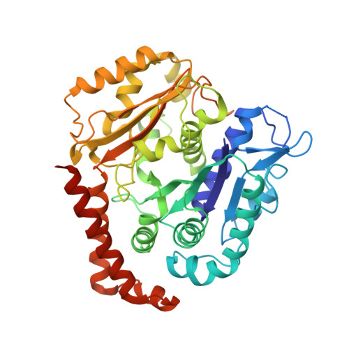
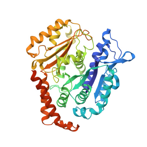
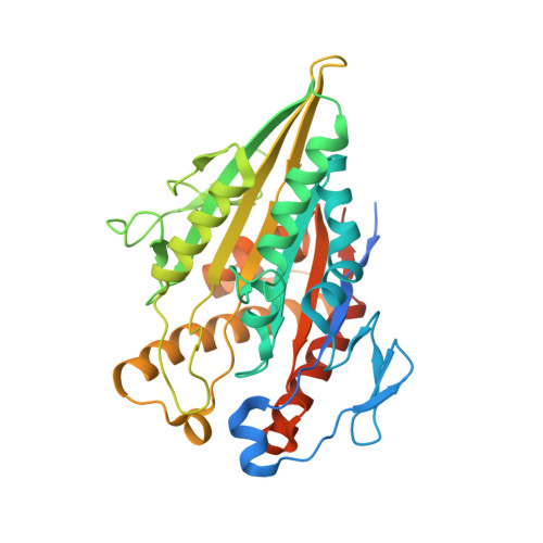
Cryo-EM of alpha-tubulin isotype-containing microtubules revealed a contracted structure of alpha 4A/ beta 2A microtubules.

Diao, L.Zheng, W.Zhao, Q.Liu, M.Fu, Z.Zhang, X.Bao, L.Cong, Y.

(2023) Acta Biochim Biophys Sin (Shanghai) 55: 1551-1560

  • DOI: https://doi.org/10.3724/abbs.2023130
  • Primary Citation of Related Structures:  
    8IXA, 8IXB, 8IXD, 8IXE, 8IXF, 8IXG

  • PubMed Abstract: 

    Microtubules are hollow α/β-tubulin heterodimeric polymers that play critical roles in cells. In vertebrates, both α- and β-tubulins have multiple isotypes encoded by different genes, which are intrinsic factors in regulating microtubule functions. However, the structures of microtubules composed of different tubulin isotypes, especially α-tubulin isotypes, remain largely unknown. Here, we purify recombinant tubulin heterodimers composed of different mouse α-tubulin isotypes, including α1A, α1C and α4A, with the β-tubulin isotype β2A. We further assemble and determine the cryo-electron microscopy (cryo-EM) structures of α1A/β2A, α1C/β2A, and α4A/β2A microtubules. Our structural analysis demonstrates that α4A/β2A microtubules exhibit longitudinal contraction between tubulin interdimers compared with α1A/β2A and α1C/β2A microtubules. Collectively, our findings reveal that α-tubulin isotype composition can tune microtubule structures, and also provide evidence for the "tubulin code" hypothesis.


  • Organizational Affiliation

    State Key Laboratory of Cell Biology, Shanghai Institute of Biochemistry and Cell Biology, Center for Excellence in Molecular Cell Science, University of Chinese Academy of Sciences, Chinese Academy of Sciences, Shanghai 200031, China.


Macromolecules
Find similar proteins by:  (by identity cutoff)  |  3D Structure
Entity ID: 1
MoleculeChains Sequence LengthOrganismDetailsImage
Tubulin alpha-1A chainA [auth H],
D [auth A],
G [auth E],
J [auth F]
457Mus musculusMutation(s): 0 
Gene Names: Tuba1aTuba1
EC: 3.6.5
UniProt & NIH Common Fund Data Resources
Find proteins for P68369 (Mus musculus)
Explore P68369 
Go to UniProtKB:  P68369
IMPC:  MGI:98869
Entity Groups  
Sequence Clusters30% Identity50% Identity70% Identity90% Identity95% Identity100% Identity
UniProt GroupP68369
Sequence Annotations
Expand
  • Reference Sequence
Find similar proteins by:  (by identity cutoff)  |  3D Structure
Entity ID: 2
MoleculeChains Sequence LengthOrganismDetailsImage
Tubulin beta-2A chainB [auth W],
E [auth Q],
H [auth U],
K [auth V]
457Mus musculusMutation(s): 0 
Gene Names: Tubb2aTubb2
UniProt & NIH Common Fund Data Resources
Find proteins for Q7TMM9 (Mus musculus)
Explore Q7TMM9 
Go to UniProtKB:  Q7TMM9
IMPC:  MGI:107861
Entity Groups  
Sequence Clusters30% Identity50% Identity70% Identity90% Identity95% Identity100% Identity
UniProt GroupQ7TMM9
Sequence Annotations
Expand
  • Reference Sequence
Find similar proteins by:  (by identity cutoff)  |  3D Structure
Entity ID: 3
MoleculeChains Sequence LengthOrganismDetailsImage
Kinesin-1 heavy chainC [auth l],
F [auth g],
I [auth k],
L [auth m]
372Homo sapiensMutation(s): 0 
Gene Names: KIF5BKNSKNS1
UniProt & NIH Common Fund Data Resources
Find proteins for P33176 (Homo sapiens)
Explore P33176 
Go to UniProtKB:  P33176
PHAROS:  P33176
GTEx:  ENSG00000170759 
Entity Groups  
Sequence Clusters30% Identity50% Identity70% Identity90% Identity95% Identity100% Identity
UniProt GroupP33176
Sequence Annotations
Expand
  • Reference Sequence
Small Molecules
Ligands 3 Unique
IDChains Name / Formula / InChI Key2D Diagram3D Interactions
GTP
Query on GTP

Download Ideal Coordinates CCD File 
M [auth W],
P [auth Q],
S [auth U],
V
GUANOSINE-5'-TRIPHOSPHATE
C10 H16 N5 O14 P3
XKMLYUALXHKNFT-UUOKFMHZSA-N
G2P
Query on G2P

Download Ideal Coordinates CCD File 
N [auth W],
Q,
T [auth U],
W [auth V]
PHOSPHOMETHYLPHOSPHONIC ACID GUANYLATE ESTER
C11 H18 N5 O13 P3
GXTIEXDFEKYVGY-KQYNXXCUSA-N
ATP
Query on ATP

Download Ideal Coordinates CCD File 
O [auth l],
R [auth g],
U [auth k],
X [auth m]
ADENOSINE-5'-TRIPHOSPHATE
C10 H16 N5 O13 P3
ZKHQWZAMYRWXGA-KQYNXXCUSA-N
Experimental Data & Validation

Experimental Data

  • Method: ELECTRON MICROSCOPY
  • Resolution: 4.20 Å
  • Aggregation State: FILAMENT 
  • Reconstruction Method: SINGLE PARTICLE 

Structure Validation

View Full Validation Report


Entry History & Funding Information

Deposition Data


Funding OrganizationLocationGrant Number
Chinese Academy of SciencesChina--

Revision History  (Full details and data files)

  • Version 1.0: 2023-08-16
    Type: Initial release
  • Version 1.1: 2023-08-30
    Changes: Database references
  • Version 1.2: 2023-10-25
    Changes: Database references