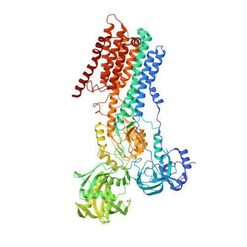
Structure and transport mechanism of the human calcium pump SPCA1.
Wu, M., Wu, C., Song, T., Pan, K., Wang, Y., Liu, Z.(2023) Cell Res 33: 533-545
- PubMed: 37258749 Search on PubMedSearch on PubMed Central
- DOI: https://doi.org/10.1038/s41422-023-00827-x
- Primary Citation Related Structures:
8IWP, 8IWR, 8IWS, 8IWT, 8IWU, 8IWW - PubMed Abstract:
Secretory-pathway Ca 2+ -ATPases (SPCAs) play critical roles in maintaining Ca 2+ homeostasis, but the exact mechanism of SPCAs-mediated Ca 2+ transport remains unclear. Here, we determined six cryo-electron microscopy (cryo-EM) structures of human SPCA1 (hSPCA1) in a series of intermediate states, revealing a near-complete conformational cycle. With the aid of molecular dynamics simulations, these structures offer a clear structural basis for Ca 2+ entry and release in hSPCA1. We found that hSPCA1 undergoes unique conformational changes during ATP binding and phosphorylation compared to other well-studied P-type II ATPases. In addition, we observed a conformational distortion of the Ca 2+ -binding site induced by the separation of transmembrane helices 4L and 6, unveiling a distinct Ca 2+ release mechanism. Particularly, we determined a structure of the long-sought CaE2P state of P-type IIA ATPases, providing valuable insights into the Ca 2+ transport cycle. Together, these findings enhance our understanding of Ca 2+ transport by hSPCA1 and broaden our knowledge of P-type ATPases.
- Department of Immunology and Microbiology, School of Life Sciences, Southern University of Science and Technology, Shenzhen, Guangdong, China.
Organizational Affiliation: