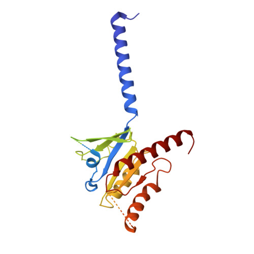
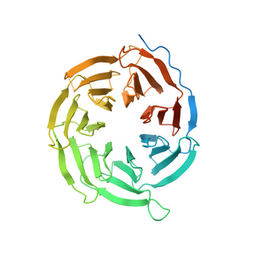
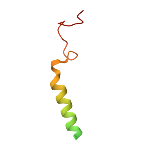
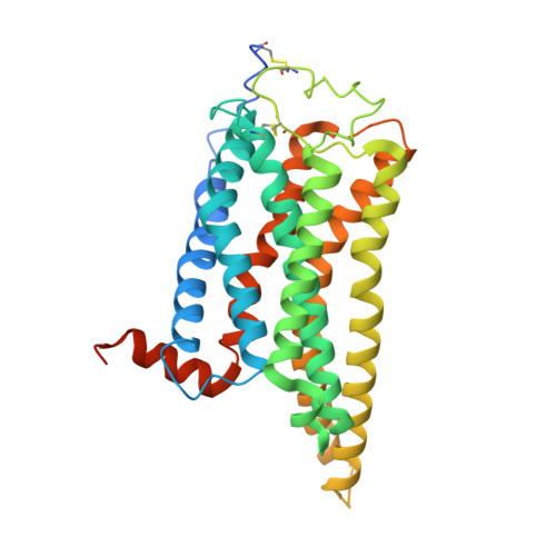
Odorants are detected as smell in the nasal epithelium of mammals by two G-protein-coupled receptor families, the odorant receptors and the trace amine-associated receptors 1,2 (TAARs). TAARs emerged following the divergence of jawed and jawless fish, and comprise a large monophyletic family of receptors that recognize volatile amine odorants to elicit both intraspecific and interspecific innate behaviours such as attraction and aversion 3-5 . Here we report cryo-electron microscopy structures of mouse TAAR9 (mTAAR9) and mTAAR9-G s or mTAAR9-G olf trimers in complex with β-phenylethylamine, N,N-dimethylcyclohexylamine or spermidine. The mTAAR9 structures contain a deep and tight ligand-binding pocket decorated with a conserved D 3.32 W 6.48 Y 7.43 motif, which is essential for amine odorant recognition. In the mTAAR9 structure, a unique disulfide bond connecting the N terminus to ECL2 is required for agonist-induced receptor activation. We identify key structural motifs of TAAR family members for detecting monoamines and polyamines and the shared sequence of different TAAR members that are responsible for recognition of the same odour chemical. We elucidate the molecular basis of mTAAR9 coupling to G s and G olf by structural characterization and mutational analysis. Collectively, our results provide a structural basis for odorant detection, receptor activation and G olf coupling of an amine olfactory receptor.
Organizational Affiliation:
Advanced Medical Research Institute, Meili Lake Translational Research Park, Cheeloo College of Medicine, Shandong University, Jinan, China.
Department of Biochemistry and Molecular Biology, Shandong University School of Medicine, Jinan, China.
Key Laboratory Experimental Teratology of the Ministry of Education and Department of Physiology, School of Basic Medical Sciences, Shandong University, Jinan, China.
Department of Physiology and Pathophysiology, School of Basic Medical Sciences, Peking University, Key Laboratory of Molecular Cardiovascular Science, Ministry of Education, Beijing, China.
Department of General Surgery, Qilu Hospital of Shandong University, Jinan, China.
Songjiang Institute and Songjiang Hospital, Shanghai Jiao Tong University School of Medicine, Shanghai, China.
Center for Brain Science, Shanghai Children's Medical Center, School of Medicine, Shanghai Jiao Tong University, Shanghai, China.
Department of Anatomy and Physiology, Ministry of Education-Shanghai Key Laboratory of Children's Environmental Health in Xinhua Hospital, Shanghai Jiao Tong University School of Medicine, Shanghai, China.
Department of Otolaryngology, Shandong Provincial Hospital Affiliated to Shandong First Medical University, Shandong, China.
MOE Key Laboratory for Nonequilibrium Synthesis and Modulation of Condensed Matter, School of Physics, Xi'an Jiaotong University, Xi'an, China.
Howard Hughes Medical Institute, Department of Cell Biology, Harvard Medical School, Boston, MA, USA.
Department of General Surgery, Qilu Hospital of Shandong University, Jinan, China. xuyunfei1988@126.com.
Advanced Medical Research Institute, Meili Lake Translational Research Park, Cheeloo College of Medicine, Shandong University, Jinan, China. yangfan1357@163.com.
Department of Biochemistry and Molecular Biology, Shandong University School of Medicine, Jinan, China. yangfan1357@163.com.
Songjiang Institute and Songjiang Hospital, Shanghai Jiao Tong University School of Medicine, Shanghai, China. liqian@shsmu.edu.cn.
Center for Brain Science, Shanghai Children's Medical Center, School of Medicine, Shanghai Jiao Tong University, Shanghai, China. liqian@shsmu.edu.cn.
Department of Anatomy and Physiology, Ministry of Education-Shanghai Key Laboratory of Children's Environmental Health in Xinhua Hospital, Shanghai Jiao Tong University School of Medicine, Shanghai, China. liqian@shsmu.edu.cn.
Shanghai Research Center for Brain Science and Brain-Inspired Intelligence, Shanghai, China. liqian@shsmu.edu.cn.
Advanced Medical Research Institute, Meili Lake Translational Research Park, Cheeloo College of Medicine, Shandong University, Jinan, China. sunjinpeng@sdu.edu.cn.
Department of Biochemistry and Molecular Biology, Shandong University School of Medicine, Jinan, China. sunjinpeng@sdu.edu.cn.
Department of Physiology and Pathophysiology, School of Basic Medical Sciences, Peking University, Key Laboratory of Molecular Cardiovascular Science, Ministry of Education, Beijing, China. sunjinpeng@sdu.edu.cn.