


Discovery and Exploration of Monosaccharide Linked Dimers to Target Fibrosis
Swidorski, J.J., Beno, B.B., Liu, C., Yoon, D., Ghosh, K., Sale, H., Shah, D., Acharya, K., Yanchunas, J., Ellsworth, B., Cheng, D., Regueiro-Ren, A.To be published.
Experimental Data Snapshot
Starting Model: experimental
View more details
Entity ID: 1 | |||||
|---|---|---|---|---|---|
| Molecule | Chains | Sequence Length | Organism | Details | Image |
| Galectin-3 | 182 | Homo sapiens | Mutation(s): 0 Gene Names: LGALS3, MAC2 | 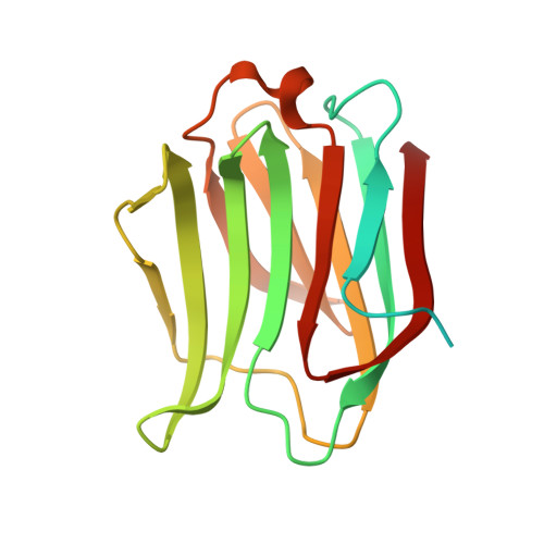 | |
UniProt & NIH Common Fund Data Resources | |||||
PHAROS: P17931 GTEx: ENSG00000131981 | |||||
Entity Groups | |||||
| Sequence Clusters | 30% Identity50% Identity70% Identity90% Identity95% Identity100% Identity | ||||
| UniProt Group | P17931 | ||||
Sequence AnnotationsExpand | |||||
Reference Sequence | |||||
| Ligands 3 Unique | |||||
|---|---|---|---|---|---|
| ID | Chains | Name / Formula / InChI Key | 2D Diagram | 3D Interactions | |
| SXP (Subject of Investigation/LOI) Download:Ideal Coordinates CCD File | D [auth A], G [auth B] | 2-[(2S,3R,4S,5R,6R)-2-[2-[2,5-bis(chloranyl)phenyl]-5-methyl-1,2,4-triazol-3-yl]-4-[4-[4-chloranyl-3,5-bis(fluoranyl)phenyl]-1,2,3-triazol-1-yl]-6-(hydroxymethyl)-5-oxidanyl-oxan-3-yl]oxyethanoic acid C25 H21 Cl3 F2 N6 O6 JCKIKNUEOCOAIU-VLZGJKPMSA-N |  | ||
| PZE (Subject of Investigation/LOI) Download:Ideal Coordinates CCD File | E [auth A] | piperazine C4 H10 N2 GLUUGHFHXGJENI-UHFFFAOYSA-N |  | ||
| CL Download:Ideal Coordinates CCD File | C [auth A], F [auth B] | CHLORIDE ION Cl VEXZGXHMUGYJMC-UHFFFAOYSA-M |  | ||
| Length ( Å ) | Angle ( ˚ ) |
|---|---|
| a = 34.35 | α = 111.12 |
| b = 43.34 | β = 105.87 |
| c = 48.15 | γ = 90.42 |
| Software Name | Purpose |
|---|---|
| REFMAC | refinement |
| XDS | data reduction |
| Aimless | data scaling |
| PHASER | phasing |
| Funding Organization | Location | Grant Number |
|---|---|---|
| Other private | -- |


