8IHH

Cryo-EM structure of HCA2-Gi complex with LUF6283


Experimental Data Snapshot

  • Method: ELECTRON MICROSCOPY
  • Resolution: 3.06 Å
  • Aggregation State: PARTICLE 
  • Reconstruction Method: SINGLE PARTICLE 

wwPDB Validation   3D Report Full Report


This is version 1.1 of the entry. See complete history


Literature

Structural basis of hydroxycarboxylic acid receptor signaling mechanisms through ligand binding.

Suzuki, S.Tanaka, K.Nishikawa, K.Suzuki, H.Oshima, A.Fujiyoshi, Y.

(2023) Nat Commun 14: 5899-5899

  • DOI: https://doi.org/10.1038/s41467-023-41650-7
  • Primary Citation of Related Structures:  
    8IHB, 8IHF, 8IHH, 8IHI, 8IHJ, 8IHK

  • PubMed Abstract: 

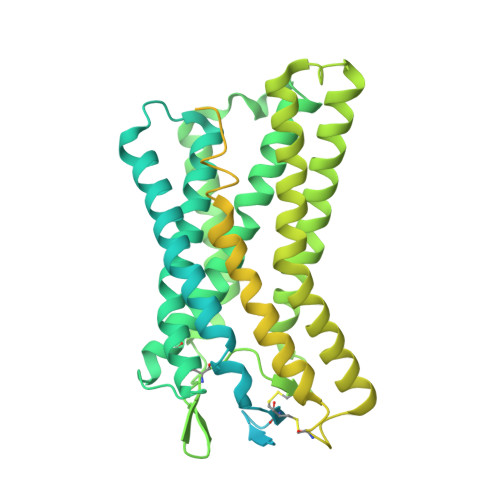
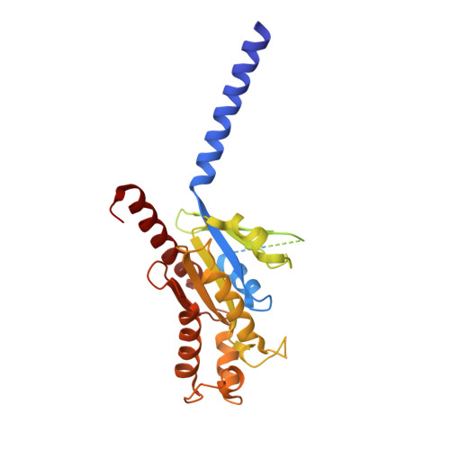
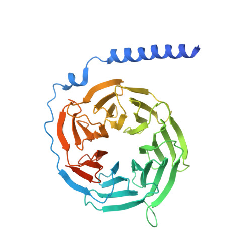
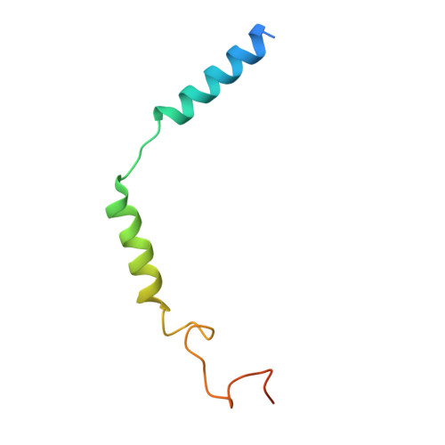
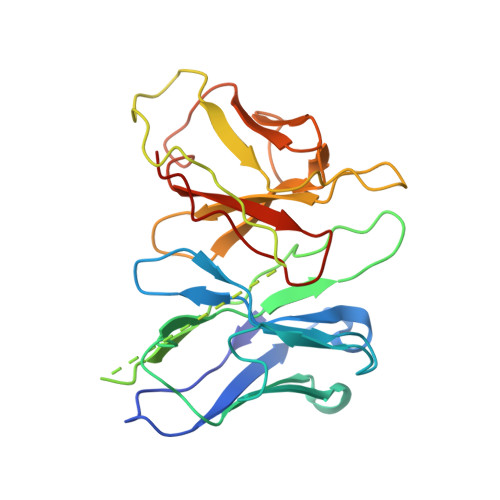
    Hydroxycarboxylic acid receptors (HCA) are expressed in various tissues and immune cells. HCA2 and its agonist are thus important targets for treating inflammatory and metabolic disorders. Only limited information is available, however, on the active-state binding of HCAs with agonists. Here, we present cryo-EM structures of human HCA2-Gi and HCA3-Gi signaling complexes binding with multiple compounds bound. Agonists were revealed to form a salt bridge with arginine, which is conserved in the HCA family, to activate these receptors. Extracellular regions of the receptors form a lid-like structure that covers the ligand-binding pocket. Although transmembrane (TM) 6 in HCAs undergoes dynamic conformational changes, ligands do not directly interact with amino acids in TM6, suggesting that indirect signaling induces a slight shift in TM6 to activate Gi proteins. Structural analyses of agonist-bound HCA2 and HCA3 together with mutagenesis and molecular dynamics simulation provide molecular insights into HCA ligand recognition and activation mechanisms.


  • Organizational Affiliation

    TMDU Advanced Research Institute, Tokyo Medical and Dental University Bunkyo-ku, Tokyo, Japan.


Macromolecules
Find similar proteins by:  (by identity cutoff)  |  3D Structure
Entity ID: 1
MoleculeChains Sequence LengthOrganismDetailsImage
Soluble cytochrome b562,Hydroxycarboxylic acid receptor 2A [auth R]658Escherichia coliHomo sapiens
This entity is chimeric
Mutation(s): 2 
Gene Names: cybCHCAR2GPR109AHCA2HM74ANIACR1
Membrane Entity: Yes 
UniProt & NIH Common Fund Data Resources
Find proteins for P0ABE7 (Escherichia coli)
Explore P0ABE7 
Go to UniProtKB:  P0ABE7
Find proteins for Q8TDS4 (Homo sapiens)
Explore Q8TDS4 
Go to UniProtKB:  Q8TDS4
PHAROS:  Q8TDS4
GTEx:  ENSG00000182782 
Entity Groups  
Sequence Clusters30% Identity50% Identity70% Identity90% Identity95% Identity100% Identity
UniProt GroupsP0ABE7Q8TDS4
Sequence Annotations
Expand
  • Reference Sequence
Find similar proteins by:  (by identity cutoff)  |  3D Structure
Entity ID: 2
MoleculeChains Sequence LengthOrganismDetailsImage
Guanine nucleotide-binding protein G(i) subunit alpha-1B [auth A]354Homo sapiensMutation(s): 2 
Gene Names: GNAI1
Membrane Entity: Yes 
UniProt & NIH Common Fund Data Resources
Find proteins for P63096 (Homo sapiens)
Explore P63096 
Go to UniProtKB:  P63096
PHAROS:  P63096
GTEx:  ENSG00000127955 
Entity Groups  
Sequence Clusters30% Identity50% Identity70% Identity90% Identity95% Identity100% Identity
UniProt GroupP63096
Sequence Annotations
Expand
  • Reference Sequence
Find similar proteins by:  (by identity cutoff)  |  3D Structure
Entity ID: 3
MoleculeChains Sequence LengthOrganismDetailsImage
Guanine nucleotide-binding protein G(I)/G(S)/G(T) subunit beta-1C [auth B]382Mus musculusMutation(s): 0 
Gene Names: Gnb1
UniProt & NIH Common Fund Data Resources
Find proteins for P62874 (Mus musculus)
Explore P62874 
Go to UniProtKB:  P62874
IMPC:  MGI:95781
Entity Groups  
Sequence Clusters30% Identity50% Identity70% Identity90% Identity95% Identity100% Identity
UniProt GroupP62874
Sequence Annotations
Expand
  • Reference Sequence
Find similar proteins by:  (by identity cutoff)  |  3D Structure
Entity ID: 4
MoleculeChains Sequence LengthOrganismDetailsImage
Guanine nucleotide-binding protein G(I)/G(S)/G(O) subunit gamma-2D [auth C]70Mus musculusMutation(s): 0 
Gene Names: Gng2
Membrane Entity: Yes 
UniProt
Find proteins for P63213 (Mus musculus)
Explore P63213 
Go to UniProtKB:  P63213
Entity Groups  
Sequence Clusters30% Identity50% Identity70% Identity90% Identity95% Identity100% Identity
UniProt GroupP63213
Sequence Annotations
Expand
  • Reference Sequence
Find similar proteins by:  (by identity cutoff)  |  3D Structure
Entity ID: 5
MoleculeChains Sequence LengthOrganismDetailsImage
scFv16E [auth S]248synthetic constructMutation(s): 0 
Entity Groups  
Sequence Clusters30% Identity50% Identity70% Identity90% Identity95% Identity100% Identity
Sequence Annotations
Expand
  • Reference Sequence
Small Molecules
Ligands 2 Unique
IDChains Name / Formula / InChI Key2D Diagram3D Interactions
NAG
Query on NAG

Download Ideal Coordinates CCD File 
F [auth R]2-acetamido-2-deoxy-beta-D-glucopyranose
C8 H15 N O6
OVRNDRQMDRJTHS-FMDGEEDCSA-N
P8A (Subject of Investigation/LOI)
Query on P8A

Download Ideal Coordinates CCD File 
G [auth R]5-butyl-1~{H}-pyrazole-3-carboxylic acid
C8 H12 N2 O2
ZJTXSGLJNBAMJS-UHFFFAOYSA-N
Experimental Data & Validation

Experimental Data

  • Method: ELECTRON MICROSCOPY
  • Resolution: 3.06 Å
  • Aggregation State: PARTICLE 
  • Reconstruction Method: SINGLE PARTICLE 

Structure Validation

View Full Validation Report


Entry History & Funding Information

Deposition Data


Funding OrganizationLocationGrant Number
Japan Society for the Promotion of Science (JSPS)Japan20H00451

Revision History  (Full details and data files)

  • Version 1.0: 2023-08-30
    Type: Initial release
  • Version 1.1: 2023-12-06
    Changes: Database references