8IF7 | pdb_00008if7

Crystal structure of CmnB


Experimental Data Snapshot

  • Method: X-RAY DIFFRACTION
  • Resolution: 2.20 Å
  • R-Value Free: 
    0.225 (Depositor), 0.232 (DCC) 
  • R-Value Work: 
    0.181 (Depositor), 0.188 (DCC) 
  • R-Value Observed: 
    0.183 (Depositor) 

Starting Model: in silico
View more details

wwPDB Validation 3D Report Full Report

Validation slider image for 8IF7

Ligand Structure Quality Assessment 


This is version 1.1 of the entry. See complete history

Literature

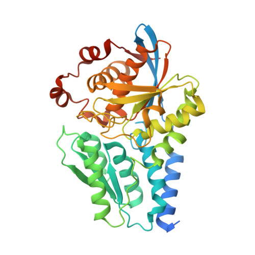
Crystal structure of CmnB involved in the biosynthesis of the nonproteinogenic amino acid L-2,3-diaminopropionic acid.

Toh, S.I.Lo, C.L.Chang, C.Y.

(2023) Acta Crystallogr F Struct Biol Commun 79: 193-199

  • DOI: https://doi.org/10.1107/S2053230X23005769
  • Primary Citation Related Structures: 
    8IF7

  • PubMed Abstract: 

    L-2,3-Diaminopropionic acid (L-Dap) is a nonproteinogenic amino acid that plays as an important role as a building block in the biosynthesis of several natural products, including capreomycin, viomycin, zwittermicin, staphyloferrin and dapdiamide. A previous study reported that CmnB and CmnK are two enzymes that are involved in the formation of L-Dap in the biosynthesis of capreomycin. CmnB catalyzes the condensation reaction of O-phospho-L-serine and L-glutamic acid to generate N-(1-amino-1-carboxyl-2-ethyl)glutamic acid, which subsequently undergoes oxidative hydrolysis via CmnK to generate the product L-Dap. Here, the crystal structure of CmnB in complex with the reaction intermediate PLP-α-aminoacrylate is reported at 2.2 Å resolution. Notably, CmnB is the second known example of a PLP-dependent enzyme that forms a monomeric structure in crystal packing. The crystal structure of CmnB also provides insights into the catalytic mechanism of the enzyme and supports the biosynthetic pathway of L-Dap reported in previous studies.


  • Organizational Affiliation
    • Department of Biological Science and Technology, National Yang Ming Chiao Tung University, Hsinchu 30010, Taiwan.

Macromolecule Content 

  • Total Structure Weight: 40.77 kDa 
  • Atom Count: 2,619 
  • Modeled Residue Count: 342 
  • Deposited Residue Count: 383 
  • Unique protein chains: 1

Macromolecules

Find similar proteins by:|  3D Structure
Entity ID: 1
MoleculeChains  Sequence LengthOrganismDetailsImage
CmnB383Saccharothrix mutabilis subsp. capreolusMutation(s): 0 
Gene Names: cmnB
UniProt
Find proteins for A6YEH3 (Saccharothrix mutabilis subsp. capreolus)
Explore A6YEH3 
Go to UniProtKB:  A6YEH3
Entity Groups
Sequence Clusters30% Identity50% Identity70% Identity90% Identity95% Identity100% Identity
UniProt GroupA6YEH3
Sequence Annotations
Expand
Reference Sequence

Small Molecules

Ligands 1 Unique
IDChains Name / Formula / InChI Key2D Diagram3D Interactions
P1T
(Subject of Investigation/LOI)

Query on P1T


Download:Ideal Coordinates CCD File
B [auth A]2-[({3-HYDROXY-2-METHYL-5-[(PHOSPHONOOXY)METHYL]PYRIDIN-4-YL}METHYL)AMINO]ACRYLIC ACID
C11 H15 N2 O7 P
BXUDKFHCAMQSRX-UHFFFAOYSA-N

Experimental Data & Validation

Experimental Data

  • Method: X-RAY DIFFRACTION
  • Resolution: 2.20 Å
  • R-Value Free:  0.225 (Depositor), 0.232 (DCC) 
  • R-Value Work:  0.181 (Depositor), 0.188 (DCC) 
  • R-Value Observed: 0.183 (Depositor) 
Space Group: P 43
Unit Cell:
Length ( Å )Angle ( ˚ )
a = 64.649α = 90
b = 64.649β = 90
c = 84.189γ = 90
Software Package:
Software NamePurpose
REFMACrefinement
SCALAdata scaling
HKL-2000data reduction
MOLREPphasing

Structure Validation

View Full Validation Report


Ligand Structure Quality Assessment 


Entry History 

& Funding Information

Deposition Data


Funding OrganizationLocationGrant Number
Ministry of Science and Technology (MoST, Taiwan)Taiwan110-2113-M-A49-026-MY3

Revision History  (Full details and data files)

  • Version 1.0: 2023-07-26
    Type: Initial release
  • Version 1.1: 2024-10-23
    Changes: Data collection, Structure summary