Structural and Biochemical Analysis of 3-Dehydroquinate Dehydratase from Corynebacterium glutamicum .
Lee, C.H., Kim, S., Seo, H., Kim, K.J.(2023) J Microbiol Biotechnol 33: 1595-1605
- PubMed: 38151830 Search on PubMedSearch on PubMed Central
- DOI: https://doi.org/10.4014/jmb.2305.05018
- Primary Citation Related Structures:
8IDR, 8IDU - PubMed Abstract:
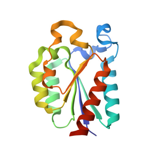
Dehydroquinate dehydratase (DHQD) catalyzes the conversion of 3-dehydroquinic acid (DHQ) into 3-dehydroshikimic acid in the mid stage of the shikimate pathway, which is essential for the biosynthesis of aromatic amino acids and folates. Here, we report two the crystal structures of type II DHQD ( Cg DHQD) derived from Corynebacterium glutamicum , which is a widely used industrial platform organism. We determined the structures for Cg DHQD WT with the citrate at a resolution of 1.80Å and Cg DHQD R19A with DHQ complexed forms at a resolution of 2.00 Å, respectively. The enzyme forms a homododecamer consisting of four trimers with three interfacial active sites. We identified the DHQ-binding site of Cg DHQD and observed an unusual binding mode of citrate inhibitor in the site with a half-opened lid loop. A structural comparison of Cg DHQD with a homolog derived from Streptomyces coelicolor revealed differences in the terminal regions, lid loop, and active site. Particularly, Cg DHQD, including some Corynebacterium species, possesses a distinctive residue P105, which is not conserved in other DHQDs at the position near the 5-hydroxyl group of DHQ. Replacements of P105 with isoleucine and valine, conserved in other DHQDs, caused an approximately 70% decrease in the activity, but replacement of S103 with threonine ( Cg DHQD S103T ) caused a 10% increase in the activity. Our biochemical studies revealed the importance of key residues and enzyme kinetics for wild type and Cg DHQD S103T , explaining the effect of the variation. This structural and biochemical study provides valuable information for understanding the reaction efficiency that varies due to structural differences caused by the unique sequences of Cg DHQD.
- School of Life Sciences, BK21 FOUR KNU Creative BioResearch Group, Kyungpook National University, Daegu 41566, Republic of Korea.
Organizational Affiliation: