8IDF

Crystal structure of human TUT1 complexed with U6 snRNA


Experimental Data Snapshot

  • Method: X-RAY DIFFRACTION
  • Resolution: 3.70 Å
  • R-Value Free: 0.339 
  • R-Value Work: 0.305 
  • R-Value Observed: 0.306 

wwPDB Validation   3D Report Full Report


This is version 1.1 of the entry. See complete history


Literature

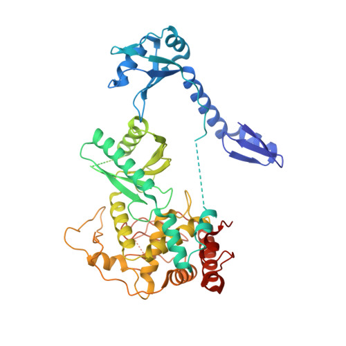
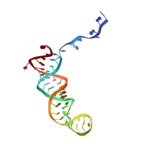
Mechanism of U6 snRNA oligouridylation by human TUT1.

Yamashita, S.Tomita, K.

(2023) Nat Commun 14: 4686-4686

  • DOI: https://doi.org/10.1038/s41467-023-40420-9
  • Primary Citation of Related Structures:  
    8IDF

  • PubMed Abstract: 

    U6 snRNA is a catalytic RNA responsible for pre-mRNA splicing reactions and undergoes various post-transcriptional modifications during its maturation process. The 3'-oligouridylation of U6 snRNA by the terminal uridylyltransferase, TUT1, provides the Lsm-binding site in U6 snRNA for U4/U6 di-snRNP formation and this ensures pre-mRNA splicing. Here, we present the crystal structure of human TUT1 (hTUT1) complexed with U6 snRNA, representing the post-uridylation of U6 snRNA by hTUT1. The N-terminal ZF-RRM and catalytic palm clamp the single-stranded AUA motif between the 5'-short stem and the 3'-telestem of U6 snRNA, and the ZF-RRM specifically recognizes the AUA motif. The ZF and the fingers hold the telestem, and the 3'-end of U6 snRNA is placed in the catalytic pocket of the palm for oligouridylation. The oligouridylation of U6 snRNA depends on the internal four-adenosine tract in the 5'-part of the telestem of U6 snRNA, and hTUT1 adds uridines until the internal adenosine tract can form base-pairs with the 3'-oligouridine tract. Together, the recognition of the specific structure and sequence of U6 snRNA by the multi-domain TUT1 protein and the intrinsic sequence and structure of U6 snRNA ensure the oligouridylation of U6 snRNA.


  • Organizational Affiliation

    Department of Computational Biology and Medical Sciences, Graduate School of Frontier Sciences, The University of Tokyo, Kashiwa, Chiba, 277-8562, Japan.


Macromolecules

Find similar proteins by:  (by identity cutoff)  |  3D Structure
Entity ID: 1
MoleculeChains Sequence LengthOrganismDetailsImage
Speckle targeted PIP5K1A-regulated poly(A) polymerase537Homo sapiensMutation(s): 8 
Gene Names: TUT1RBM21
EC: 2.7.7.19 (PDB Primary Data), 2.7.7.52 (PDB Primary Data)
UniProt & NIH Common Fund Data Resources
Find proteins for Q9H6E5 (Homo sapiens)
Explore Q9H6E5 
Go to UniProtKB:  Q9H6E5
PHAROS:  Q9H6E5
GTEx:  ENSG00000149016 
Entity Groups  
Sequence Clusters30% Identity50% Identity70% Identity90% Identity95% Identity100% Identity
UniProt GroupQ9H6E5
Sequence Annotations
Expand
  • Reference Sequence
Find similar nucleic acids by:  (by identity cutoff)  |  3D Structure
Entity ID: 2
MoleculeChains LengthOrganismImage
RNA (53-MER)53Homo sapiens
Sequence Annotations
Expand
  • Reference Sequence
Small Molecules
Ligands 1 Unique
IDChains Name / Formula / InChI Key2D Diagram3D Interactions
ZN (Subject of Investigation/LOI)
Query on ZN

Download Ideal Coordinates CCD File 
C [auth A]ZINC ION
Zn
PTFCDOFLOPIGGS-UHFFFAOYSA-N
Experimental Data & Validation

Experimental Data

  • Method: X-RAY DIFFRACTION
  • Resolution: 3.70 Å
  • R-Value Free: 0.339 
  • R-Value Work: 0.305 
  • R-Value Observed: 0.306 
  • Space Group: P 65 2 2
Unit Cell:
Length ( Å )Angle ( ˚ )
a = 78.724α = 90
b = 78.724β = 90
c = 370.272γ = 120
Software Package:
Software NamePurpose
PHENIXrefinement
XSCALEdata scaling
PDB_EXTRACTdata extraction
XDSdata reduction
PHASERphasing

Structure Validation

View Full Validation Report


Entry History & Funding Information

Deposition Data


Funding OrganizationLocationGrant Number
Japan Society for the Promotion of Science (JSPS)Japan18H03980
Japan Society for the Promotion of Science (JSPS)Japan19K16053

Revision History  (Full details and data files)

  • Version 1.0: 2023-08-09
    Type: Initial release
  • Version 1.1: 2023-08-23
    Changes: Data collection, Database references