Substrate recognition mode of a glycoside hydrolase family 42 beta-galactosidase from Bifidobacterium longum subspecies infantis ( Bi Bga42A) revealed by crystallographic and mutational analyses.
Gotoh, A., Hidaka, M., Sakurama, H., Nishimoto, M., Kitaoka, M., Sakanaka, M., Fushinobu, S., Katayama, T.(2023) Microbiome Res Rep 2: 20-20
- PubMed: 38046823 Search on PubMedSearch on PubMed Central
- DOI: https://doi.org/10.20517/mrr.2023.14
- Primary Citation Related Structures:
8IBR, 8IBS, 8IBT - PubMed Abstract:
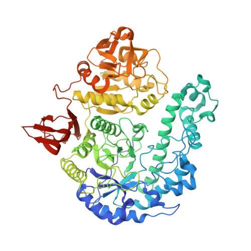
Aim: Bifidobacterium longum subsp. infantis uses a glycoside hydrolase (GH) family 42 β-galactosidase ( Bi Bga42A) for hydrolyzing lacto- N -tetraose (LNT), which is the most abundant core structure of human milk oligosaccharides (HMOs). As such, Bi Bga42A represents one of the pivotal enzymes underpinning the symbiosis between bifidobacteria and breastfed infants. Despite its importance, the structural basis underlying LNT hydrolysis by Bi Bga42A is not understood. Moreover, no substrate-complexed structures are available to date for GH42 family members. Methods: X-ray crystallography was used to determine the structures of Bi Bga42A in the apo- and liganded forms. The roles of the amino acid residues that were presumed to be involved in catalysis and substrate recognition were examined by a mutational study, in which kinetic parameters of each mutant were determined using 4-nitrophenyl-β-D-galactoside, lacto- N -biose I, LNT, and lacto- N -neotetraose (LNnT) as substrates. Conservation of those amino acid residues was examined among structure-determined GH42 β-galactosidases. Results: Crystal structures of the wild-type enzyme complexed with glycerol, the E160A/E318A double mutant complexed with galactose (Gal), and the E318S mutant complexed with LNT were determined at 1.7, 1.9, and 2.2 Å resolutions, respectively. The LNT molecule (excluding the Gal moiety at subsite +2) bound to the E318S mutant is recognized by an extensive hydrogen bond network and several hydrophobic interactions. The non-reducing end Gal moiety of LNT adopts a slightly distorted conformation and does not overlap well with the Gal molecule bound to the E160A/E318A mutant. Twelve of the sixteen amino acid residues responsible for LNT recognition and catalysis in Bi Bga42A are conserved among all homologs including β-1,6-1,3-galactosidase ( Bl Gal42A) from Bifidobacterium animalis subsp. lactis . Conclusion: Bl Gal42A is active on 3-β-galactobiose similarly to Bi Bga42A but is inactive on LNT. Interestingly, we found that the entrance of the catalytic pocket of Bl Gal42A is narrower than that of Bi Bga42A and seems not easily accessible from the solvent side due to the presence of two bulky amino acid side chains. The specificity difference may reflect the structural difference between the two enzymes.
- Graduate School of Biostudies, Kyoto University, Sakyo-ku, Kyoto 606-8502, Japan.
Organizational Affiliation: