Alteration of Substrate Specificity and Transglucosylation Activity of GH13_31 alpha-Glucosidase from Bacillus sp. AHU2216 through Site-Directed Mutagenesis of Asn258 on beta → alpha Loop 5.
Auiewiriyanukul, W., Saburi, W., Ota, T., Yu, J., Kato, K., Yao, M., Mori, H.(2023) Molecules 28
- PubMed: 37049872 Search on PubMedSearch on PubMed Central
- DOI: https://doi.org/10.3390/molecules28073109
- Primary Citation Related Structures:
8IBK, 8IDS - PubMed Abstract:
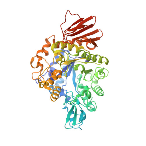
α-Glucosidase catalyzes the hydrolysis of α-d-glucosides and transglucosylation. Bacillus sp. AHU2216 α-glucosidase (BspAG13_31A), belonging to the glycoside hydrolase family 13 subfamily 31, specifically cleaves α-(1→4)-glucosidic linkages and shows high disaccharide specificity. We showed previously that the maltose moiety of maltotriose (G3) and maltotetraose (G4), covering subsites +1 and +2 of BspAG13_31A, adopts a less stable conformation than the global minimum energy conformation. This unstable d-glucosyl conformation likely arises from steric hindrance by Asn258 on β→α loop 5 of the catalytic (β/α) 8 -barrel. In this study, Asn258 mutants of BspAG13_31A were enzymatically and structurally analyzed. N258G/P mutations significantly enhanced trisaccharide specificity. The N258P mutation also enhanced the activity toward sucrose and produced erlose from sucrose through transglucosylation. N258G showed a higher specificity to transglucosylation with p -nitrophenyl α-d-glucopyranoside and maltose than the wild type. E256Q/N258G and E258Q/N258P structures in complex with G3 revealed that the maltose moiety of G3 bound at subsites +1 and +2 adopted a relaxed conformation, whereas a less stable conformation was taken in E256Q. This structural difference suggests that stabilizing the G3 conformation enhances trisaccharide specificity. The E256Q/N258G-G3 complex formed an additional hydrogen bond between Met229 and the d-glucose residue of G3 in subsite +2, and this interaction may enhance transglucosylation.
- Research Faculty of Agriculture, Hokkaido Unifversity, Sapporo 060-8589, Japan.
Organizational Affiliation: