8IB2

Structure of mammalian spectrin-actin junctional complex of membrane skeleton, Pointed-end segment, headpiece domain of dematin optimized

  • Classification: MEMBRANE PROTEIN
  • Organism(s): Sus scrofa
  • Mutation(s): No 

  • Deposited: 2023-02-09 Released: 2023-04-26 
  • Deposition Author(s): Li, N., Chen, S., Gao, N.
  • Funding Organization(s): National Science Foundation (NSF, China)

Experimental Data Snapshot

  • Method: ELECTRON MICROSCOPY
  • Resolution: 3.80 Å
  • Aggregation State: PARTICLE 
  • Reconstruction Method: SINGLE PARTICLE 

wwPDB Validation   3D Report Full Report


This is version 1.2 of the entry. See complete history


Literature

Structural basis of membrane skeleton organization in red blood cells.

Li, N.Chen, S.Xu, K.He, M.T.Dong, M.Q.Zhang, Q.C.Gao, N.

(2023) Cell 186: 1912-1929.e18

  • DOI: https://doi.org/10.1016/j.cell.2023.03.017
  • Primary Citation of Related Structures:  
    8IAH, 8IAI, 8IB2

  • PubMed Abstract: 

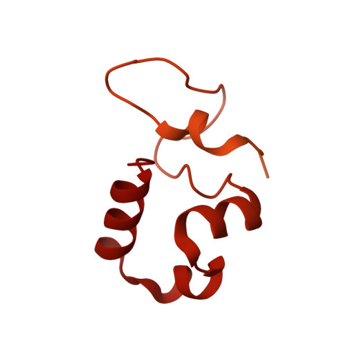
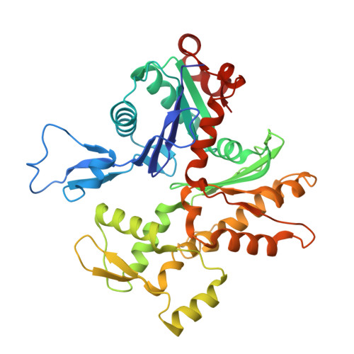
    The spectrin-based membrane skeleton is a ubiquitous membrane-associated two-dimensional cytoskeleton underneath the lipid membrane of metazoan cells. Mutations of skeleton proteins impair the mechanical strength and functions of the membrane, leading to several different types of human diseases. Here, we report the cryo-EM structures of the native spectrin-actin junctional complex (from porcine erythrocytes), which is a specialized short F-actin acting as the central organizational unit of the membrane skeleton. While an α-/β-adducin hetero-tetramer binds to the barbed end of F-actin as a flexible cap, tropomodulin and SH3BGRL2 together create an absolute cap at the pointed end. The junctional complex is strengthened by ring-like structures of dematin in the middle actin layers and by patterned periodic interactions with tropomyosin over its entire length. This work serves as a structural framework for understanding the assembly and dynamics of membrane skeleton and offers insights into mechanisms of various ubiquitous F-actin-binding factors in other F-actin systems.


  • Organizational Affiliation

    State Key Laboratory of Membrane Biology, Peking-Tsinghua Joint Center for Life Sciences, School of Life Sciences, Peking University, Beijing 100871, China; Changping Laboratory, Beijing 102206, China. Electronic address: ningningli@pku.edu.cn.


Macromolecules
Find similar proteins by:  (by identity cutoff)  |  3D Structure
Entity ID: 1
MoleculeChains Sequence LengthOrganismDetailsImage
Dematin actin binding proteinA [auth 5]405Sus scrofaMutation(s): 0 
UniProt
Find proteins for F1RMC1 (Sus scrofa)
Explore F1RMC1 
Go to UniProtKB:  F1RMC1
Entity Groups  
Sequence Clusters30% Identity50% Identity70% Identity90% Identity95% Identity100% Identity
UniProt GroupF1RMC1
Sequence Annotations
Expand
  • Reference Sequence
Find similar proteins by:  (by identity cutoff)  |  3D Structure
Entity ID: 2
MoleculeChains Sequence LengthOrganismDetailsImage
Actin, cytoplasmic 1
B, C, D, E, F
375Sus scrofaMutation(s): 0 
UniProt
Find proteins for Q6QAQ1 (Sus scrofa)
Explore Q6QAQ1 
Go to UniProtKB:  Q6QAQ1
Entity Groups  
Sequence Clusters30% Identity50% Identity70% Identity90% Identity95% Identity100% Identity
UniProt GroupQ6QAQ1
Sequence Annotations
Expand
  • Reference Sequence
Experimental Data & Validation

Experimental Data

  • Method: ELECTRON MICROSCOPY
  • Resolution: 3.80 Å
  • Aggregation State: PARTICLE 
  • Reconstruction Method: SINGLE PARTICLE 

Structure Validation

View Full Validation Report


Entry History & Funding Information

Deposition Data


Funding OrganizationLocationGrant Number
National Science Foundation (NSF, China)China--

Revision History  (Full details and data files)

  • Version 1.0: 2023-04-26
    Type: Initial release
  • Version 1.1: 2023-05-03
    Changes: Structure summary
  • Version 1.2: 2023-05-10
    Changes: Database references