8I7P

Crystal structure of Ricin A chain bound with N2-(2-amino-4-oxo-3,4-dihydropteridine-7-carbonyl)glycyl-L-tyrosine


Experimental Data Snapshot

  • Method: X-RAY DIFFRACTION
  • Resolution: 1.60 Å
  • R-Value Free: 0.232 
  • R-Value Work: 0.215 
  • R-Value Observed: 0.216 

wwPDB Validation   3D Report Full Report


This is version 1.0 of the entry. See complete history


Literature

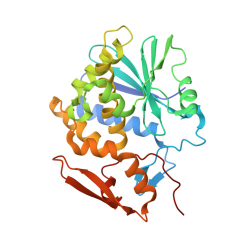
Crystal structure of ricin toxin A chain complexed with a highly potent pterin-based small-molecular inhibitor.

Goto, M.Sakamoto, N.Higashi, S.Kawata, R.Nagatsu, K.Saito, R.

(2023) J Enzyme Inhib Med Chem 38: 2219038-2219038

  • DOI: https://doi.org/10.1080/14756366.2023.2219038
  • Primary Citation of Related Structures:  
    8I7P

  • PubMed Abstract: 

    Ricin toxin A chain (RTA), from Ricinus communis , is a deadly protein that inactivates ribosomes by degrading an adenine residue at position 4324 in 28S rRNA. Recently, we have demonstrated that pterin-7-carboxamides with peptide pendants were potent RTA inhibitors. Among these, N -(pterin-7-carbonyl)glycyl-L-tyrosine ( 7PCGY ) is the most potent RTA inhibitor as a small organic molecule. However, despite this fascinating inhibitory activity, the mode of interaction of 7PCGY with RTA remains elusive. This study aimed to elucidate the factors responsible for the high RTA inhibitory activity of 7PCGY based on X-ray crystallographic analysis. Herein, we report the successfully resolved X-ray crystal structure of 7PCGY /RTA complexes, revealing that the interaction between the phenolic hydroxy group in 7PCGY and Asn78 of RTA through a hydrogen bonding and the conformational change of Tyr80 and Asn122 are responsible for the high RTA inhibitory activity of 7PCGY .


  • Organizational Affiliation

    Department of Molecular Bioscience, Toho University, Japan.


Macromolecules
Find similar proteins by:  (by identity cutoff)  |  3D Structure
Entity ID: 1
MoleculeChains Sequence LengthOrganismDetailsImage
Ricin A chain274Ricinus communisMutation(s): 0 
EC: 3.2.2.22
UniProt
Find proteins for P02879 (Ricinus communis)
Explore P02879 
Go to UniProtKB:  P02879
Entity Groups  
Sequence Clusters30% Identity50% Identity70% Identity90% Identity95% Identity100% Identity
UniProt GroupP02879
Sequence Annotations
Expand
  • Reference Sequence

Find similar proteins by:  Sequence   |   3D Structure  

Entity ID: 2
MoleculeChains Sequence LengthOrganismDetailsImage
6-(2-ethyl-4-hydroxyphenyl)-1H-indazole-3-carboxamide3synthetic constructMutation(s): 0 
Sequence Annotations
Expand
  • Reference Sequence
Experimental Data & Validation

Experimental Data

  • Method: X-RAY DIFFRACTION
  • Resolution: 1.60 Å
  • R-Value Free: 0.232 
  • R-Value Work: 0.215 
  • R-Value Observed: 0.216 
  • Space Group: P 41 21 2
Unit Cell:
Length ( Å )Angle ( ˚ )
a = 67.803α = 90
b = 67.803β = 90
c = 141.38γ = 90
Software Package:
Software NamePurpose
REFMACrefinement
SCALAdata scaling
XDSdata reduction
MOLREPphasing

Structure Validation

View Full Validation Report


Entry History & Funding Information

Deposition Data


Funding OrganizationLocationGrant Number
Japan Society for the Promotion of Science (JSPS)JapanJP19K05699

Revision History  (Full details and data files)

  • Version 1.0: 2023-09-20
    Type: Initial release