8I2F

Crystal structure of Bacillus subtilis LytE catalytic domain in complex with IseA


Experimental Data Snapshot

  • Method: X-RAY DIFFRACTION
  • Resolution: 2.03 Å
  • R-Value Free: 0.208 
  • R-Value Work: 0.173 
  • R-Value Observed: 0.175 

wwPDB Validation   3D Report Full Report


This is version 1.2 of the entry. See complete history


Literature

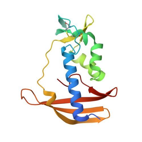
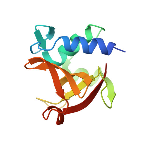
Structural insights into the regulation of peptidoglycan DL-endopeptidases by inhibitory protein IseA.

Tandukar, S.Kwon, E.Kim, D.Y.

(2023) Structure 31: 619-628.e4

  • DOI: https://doi.org/10.1016/j.str.2023.02.013
  • Primary Citation of Related Structures:  
    8I2D, 8I2E, 8I2F

  • PubMed Abstract: 

    Peptidoglycan, a physical barrier that protects bacteria from the environment, is constantly degraded and resynthesized for remodeling during cell growth and division. Because excessive or insufficient peptidoglycan hydrolysis affects bacterial homeostasis and viability, peptidoglycan degradation must be precisely regulated. In Bacillus subtilis, DL-endopeptidases play an essential role in peptidoglycan remodeling, and their activity is regulated by IseA. Here, we report the crystal structure of peptidoglycan DL-endopeptidase LytE complexed with IseA. In the crystal structure, the inhibitory loop connecting the two lobes of IseA blocks the active site of LytE by mimicking its substrate. Consistently, mutations in the inhibitory loop resulted in the loss of IseA activity. The structure also shows that conformational rearrangements in both LytE and IseA restrict access of the inhibitory loop to the LytE catalytic site. These results reveal an inhibition mechanism of peptidoglycan DL-endopeptidase in which the inhibitory protein mimics the substrate but is not degraded.


  • Organizational Affiliation

    College of Pharmacy, Yeungnam University, Gyeongsan 38541, South Korea.


Macromolecules
Find similar proteins by:  (by identity cutoff)  |  3D Structure
Entity ID: 1
MoleculeChains Sequence LengthOrganismDetailsImage
Uncharacterized protein YoeB
A, C
160Bacillus subtilis subsp. subtilis str. 168Mutation(s): 0 
Gene Names: yoeB
UniProt
Find proteins for O34841 (Bacillus subtilis (strain 168))
Explore O34841 
Go to UniProtKB:  O34841
Entity Groups  
Sequence Clusters30% Identity50% Identity70% Identity90% Identity95% Identity100% Identity
UniProt GroupO34841
Sequence Annotations
Expand
  • Reference Sequence
Find similar proteins by:  (by identity cutoff)  |  3D Structure
Entity ID: 2
MoleculeChains Sequence LengthOrganismDetailsImage
Probable peptidoglycan endopeptidase LytE
B, D
122Bacillus subtilis subsp. subtilis str. 168Mutation(s): 0 
Gene Names: lytE
EC: 3.4
UniProt
Find proteins for P54421 (Bacillus subtilis (strain 168))
Explore P54421 
Go to UniProtKB:  P54421
Entity Groups  
Sequence Clusters30% Identity50% Identity70% Identity90% Identity95% Identity100% Identity
UniProt GroupP54421
Sequence Annotations
Expand
  • Reference Sequence
Experimental Data & Validation

Experimental Data

  • Method: X-RAY DIFFRACTION
  • Resolution: 2.03 Å
  • R-Value Free: 0.208 
  • R-Value Work: 0.173 
  • R-Value Observed: 0.175 
  • Space Group: P 21 21 21
Unit Cell:
Length ( Å )Angle ( ˚ )
a = 60.83α = 90
b = 74.64β = 90
c = 153.08γ = 90
Software Package:
Software NamePurpose
PHENIXrefinement
MOSFLMdata reduction
Aimlessdata scaling
PHASERphasing

Structure Validation

View Full Validation Report


Entry History & Funding Information

Deposition Data


Funding OrganizationLocationGrant Number
Not funded--

Revision History  (Full details and data files)

  • Version 1.0: 2023-04-05
    Type: Initial release
  • Version 1.1: 2023-04-19
    Changes: Structure summary
  • Version 1.2: 2023-05-17
    Changes: Database references