8HUM

X-ray structure of human PPAR gamma ligand binding domain-lanifibranor-SRC1 coactivator peptide co-crystals obtained by co-crystallization


Experimental Data Snapshot

  • Method: X-RAY DIFFRACTION
  • Resolution: 2.29 Å
  • R-Value Free: 0.250 
  • R-Value Work: 0.206 
  • R-Value Observed: 0.208 

wwPDB Validation   3D Report Full Report


Ligand Structure Quality Assessment 


This is version 1.1 of the entry. See complete history


Literature

Functional and Structural Insights into the Human PPAR alpha / delta / gamma Targeting Preferences of Anti-NASH Investigational Drugs, Lanifibranor, Seladelpar, and Elafibranor.

Kamata, S.Honda, A.Ishikawa, R.Akahane, M.Fujita, A.Kaneko, C.Miyawaki, S.Habu, Y.Shiiyama, Y.Uchii, K.Machida, Y.Oyama, T.Ishii, I.

(2023) Antioxidants (Basel) 12

  • DOI: https://doi.org/10.3390/antiox12081523
  • Primary Citation of Related Structures:  
    8HUK, 8HUL, 8HUM, 8HUN, 8HUO, 8HUP, 8HUQ

  • PubMed Abstract: 

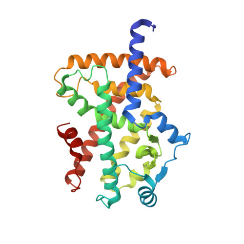
    No therapeutic drugs are currently available for nonalcoholic steatohepatitis (NASH) that progresses from nonalcoholic fatty liver via oxidative stress-involved pathways. Three cognate peroxisome proliferator-activated receptor (PPAR) subtypes (PPARα/δ/γ) are considered as attractive targets. Although lanifibranor (PPARα/δ/γ pan agonist) and saroglitazar (PPARα/γ dual agonist) are currently under investigation in clinical trials for NASH, the development of seladelpar (PPARδ-selective agonist), elafibranor (PPARα/δ dual agonist), and many other dual/pan agonists has been discontinued due to serious side effects or little/no efficacies. This study aimed to obtain functional and structural insights into the potency, efficacy, and selectivity against PPARα/δ/γ of three current and past anti-NASH investigational drugs: lanifibranor, seladelpar, and elafibranor. Ligand activities were evaluated by three assays to detect different facets of the PPAR activation: transactivation assay, coactivator recruitment assay, and thermal stability assay. Seven high-resolution cocrystal structures (namely, those of the PPARα/δ/γ-ligand-binding domain (LBD)-lanifibranor, PPARα/δ/γ-LBD-seladelpar, and PPARα-LBD-elafibranor) were obtained through X-ray diffraction analyses, six of which represent the first deposit in the Protein Data Bank. Lanifibranor and seladelpar were found to bind to different regions of the PPARα/δ/γ-ligand-binding pockets and activated all PPAR subtypes with different potencies and efficacies in the three assays. In contrast, elafibranor induced transactivation and coactivator recruitment (not thermal stability) of all PPAR subtypes, but the PPARδ/γ-LBD-elafibranor cocrystals were not obtained. These results illustrate the highly variable PPARα/δ/γ activation profiles and binding modes of these PPAR ligands that define their pharmacological actions.


  • Organizational Affiliation

    Department of Health Chemistry, Showa Pharmaceutical University, Machida 194-8543, Tokyo, Japan.


Macromolecules
Find similar proteins by:  (by identity cutoff)  |  3D Structure
Entity ID: 1
MoleculeChains Sequence LengthOrganismDetailsImage
Isoform 1 of Peroxisome proliferator-activated receptor gamma279Homo sapiensMutation(s): 0 
Gene Names: PPARG
UniProt & NIH Common Fund Data Resources
Find proteins for P37231 (Homo sapiens)
Explore P37231 
Go to UniProtKB:  P37231
PHAROS:  P37231
GTEx:  ENSG00000132170 
Entity Groups  
Sequence Clusters30% Identity50% Identity70% Identity90% Identity95% Identity100% Identity
UniProt GroupP37231
Sequence Annotations
Expand
  • Reference Sequence

Find similar proteins by:  Sequence   |   3D Structure  

Entity ID: 2
MoleculeChains Sequence LengthOrganismDetailsImage
15-meric peptide from Nuclear receptor coactivator 115Homo sapiensMutation(s): 0 
EC: 2.3.1.48
UniProt & NIH Common Fund Data Resources
Find proteins for Q15788 (Homo sapiens)
Explore Q15788 
Go to UniProtKB:  Q15788
PHAROS:  Q15788
GTEx:  ENSG00000084676 
Entity Groups  
Sequence Clusters30% Identity50% Identity70% Identity90% Identity95% Identity100% Identity
UniProt GroupQ15788
Sequence Annotations
Expand
  • Reference Sequence
Small Molecules
Ligands 1 Unique
IDChains Name / Formula / InChI Key2D Diagram3D Interactions
BJB (Subject of Investigation/LOI)
Query on BJB

Download Ideal Coordinates CCD File 
C [auth A]4-[1-(1,3-benzothiazol-6-ylsulfonyl)-5-chloro-indol-2-yl]butanoic acid
C19 H15 Cl N2 O4 S2
OQDQIFQRNZIEEJ-UHFFFAOYSA-N
Experimental Data & Validation

Experimental Data

  • Method: X-RAY DIFFRACTION
  • Resolution: 2.29 Å
  • R-Value Free: 0.250 
  • R-Value Work: 0.206 
  • R-Value Observed: 0.208 
  • Space Group: P 21 21 21
Unit Cell:
Length ( Å )Angle ( ˚ )
a = 49.862α = 90
b = 63.728β = 90
c = 123.484γ = 90
Software Package:
Software NamePurpose
XDSdata reduction
Aimlessdata scaling
PHASERphasing
PHENIXrefinement
PDB_EXTRACTdata extraction

Structure Validation

View Full Validation Report


Ligand Structure Quality Assessment 


Entry History & Funding Information

Deposition Data


Funding OrganizationLocationGrant Number
Japan Society for the Promotion of Science (JSPS)Japan22K15049
Japan Society for the Promotion of Science (JSPS)Japan22H05577
Japan Agency for Medical Research and Development (AMED)JapanJP21am0101071

Revision History  (Full details and data files)

  • Version 1.0: 2023-08-09
    Type: Initial release
  • Version 1.1: 2023-09-06
    Changes: Data collection, Database references