8HTG

Crystal structure of Golf in complex with GTP-gamma S and Mg


Experimental Data Snapshot

  • Method: X-RAY DIFFRACTION
  • Resolution: 2.91 Å
  • R-Value Free: 0.267 
  • R-Value Work: 0.230 
  • R-Value Observed: 0.232 

wwPDB Validation   3D Report Full Report


Ligand Structure Quality Assessment 


This is version 1.0 of the entry. See complete history


Literature

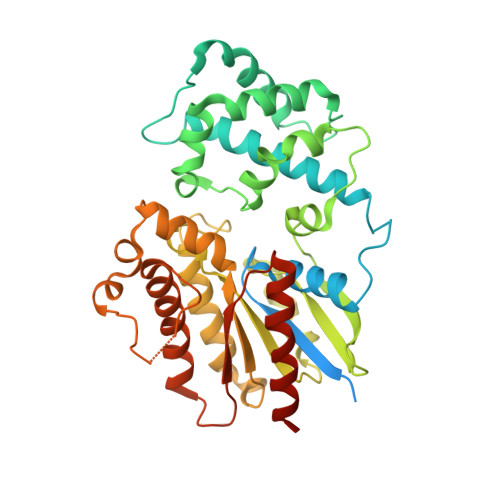
Understanding the molecular mechanisms of odorant binding and activation of the human OR52 family.

Choi, C.Bae, J.Kim, S.Lee, S.Kang, H.Kim, J.Bang, I.Kim, K.Huh, W.K.Seok, C.Park, H.Im, W.Choi, H.J.

(2023) Nat Commun 14: 8105-8105

  • DOI: https://doi.org/10.1038/s41467-023-43983-9
  • Primary Citation of Related Structures:  
    8HTG, 8HTI, 8J46, 8W77

  • PubMed Abstract: 

    Structural and mechanistic studies on human odorant receptors (ORs), key in olfactory signaling, are challenging because of their low surface expression in heterologous cells. The recent structure of OR51E2 bound to propionate provided molecular insight into odorant recognition, but the lack of an inactive OR structure limited understanding of the activation mechanism of ORs upon odorant binding. Here, we determined the cryo-electron microscopy structures of consensus OR52 (OR52 cs ), a representative of the OR52 family, in the ligand-free (apo) and octanoate-bound states. The apo structure of OR52 cs reveals a large opening between transmembrane helices (TMs) 5 and 6. A comparison between the apo and active structures of OR52 cs demonstrates the inward and outward movements of the extracellular and intracellular segments of TM6, respectively. These results, combined with molecular dynamics simulations and signaling assays, shed light on the molecular mechanisms of odorant binding and activation of the OR52 family.


  • Organizational Affiliation

    Department of Biological Sciences, Seoul National University, Seoul, 08826, Republic of Korea.


Macromolecules
Find similar proteins by:  (by identity cutoff)  |  3D Structure
Entity ID: 1
MoleculeChains Sequence LengthOrganismDetailsImage
Guanine nucleotide-binding protein G(olf) subunit alpha
A, B, C, D
395Mus musculusMutation(s): 1 
Gene Names: Gnal
UniProt
Find proteins for Q8CGK7 (Mus musculus)
Explore Q8CGK7 
Go to UniProtKB:  Q8CGK7
Entity Groups  
Sequence Clusters30% Identity50% Identity70% Identity90% Identity95% Identity100% Identity
UniProt GroupQ8CGK7
Sequence Annotations
Expand
  • Reference Sequence
Small Molecules
Ligands 4 Unique
IDChains Name / Formula / InChI Key2D Diagram3D Interactions
GSP (Subject of Investigation/LOI)
Query on GSP

Download Ideal Coordinates CCD File 
G [auth A],
L [auth B],
N [auth C],
Q [auth D]
5'-GUANOSINE-DIPHOSPHATE-MONOTHIOPHOSPHATE
C10 H16 N5 O13 P3 S
XOFLBQFBSOEHOG-UUOKFMHZSA-N
PO4
Query on PO4

Download Ideal Coordinates CCD File 
H [auth A],
I [auth A],
O [auth C]
PHOSPHATE ION
O4 P
NBIIXXVUZAFLBC-UHFFFAOYSA-K
EDO
Query on EDO

Download Ideal Coordinates CCD File 
E [auth A],
J [auth B]
1,2-ETHANEDIOL
C2 H6 O2
LYCAIKOWRPUZTN-UHFFFAOYSA-N
MG (Subject of Investigation/LOI)
Query on MG

Download Ideal Coordinates CCD File 
F [auth A],
K [auth B],
M [auth C],
P [auth D]
MAGNESIUM ION
Mg
JLVVSXFLKOJNIY-UHFFFAOYSA-N
Experimental Data & Validation

Experimental Data

  • Method: X-RAY DIFFRACTION
  • Resolution: 2.91 Å
  • R-Value Free: 0.267 
  • R-Value Work: 0.230 
  • R-Value Observed: 0.232 
  • Space Group: P 1 21 1
Unit Cell:
Length ( Å )Angle ( ˚ )
a = 121.73α = 90
b = 52.36β = 118.53
c = 125.51γ = 90
Software Package:
Software NamePurpose
PHENIXrefinement
XDSdata scaling
XDSdata reduction
PHASERphasing

Structure Validation

View Full Validation Report


Ligand Structure Quality Assessment 


Entry History & Funding Information

Deposition Data


Funding OrganizationLocationGrant Number
Samsung Science and Technology FoundationKorea, Republic OfSSTF-BA1901-09

Revision History  (Full details and data files)

  • Version 1.0: 2023-12-20
    Type: Initial release