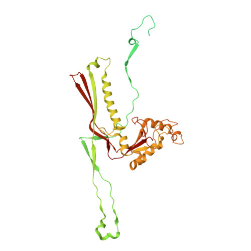
Nearly complete structure of bacteriophage DT57C reveals architecture of head-to-tail interface and lateral tail fibers.
Ayala, R., Moiseenko, A.V., Chen, T.H., Kulikov, E.E., Golomidova, A.K., Orekhov, P.S., Street, M.A., Sokolova, O.S., Letarov, A.V., Wolf, M.(2023) Nat Commun 14: 8205-8205
- PubMed: 38081816 Search on PubMedSearch on PubMed Central
- DOI: https://doi.org/10.1038/s41467-023-43824-9
- Primary Citation Related Structures:
8HO3, 8HQK, 8HQO, 8HQZ, 8HRE, 8HRG - PubMed Abstract:
The T5 family of viruses are tailed bacteriophages characterized by a long non-contractile tail. The bacteriophage DT57C is closely related to the paradigmal T5 phage, though it recognizes a different receptor (BtuB) and features highly divergent lateral tail fibers (LTF). Considerable portions of T5-like phages remain structurally uncharacterized. Here, we present the structure of DT57C determined by cryo-EM, and an atomic model of the virus, which was further explored using all-atom molecular dynamics simulations. The structure revealed a unique way of LTF attachment assisted by a dodecameric collar protein LtfC, and an unusual composition of the phage neck constructed of three protein rings. The tape measure protein (TMP) is organized within the tail tube in a three-stranded parallel α-helical coiled coil which makes direct contact with the genomic DNA. The presence of the C-terminal fragment of the TMP that remains within the tail tip suggests that the tail tip complex returns to its original state after DNA ejection. Our results provide a complete atomic structure of a T5-like phage, provide insights into the process of DNA ejection as well as a structural basis for the design of engineered phages and future mechanistic studies.
- Molecular Cryo-Electron Microscopy Unit, Okinawa Institute of Science and Technology Graduate University, 1919-1 Tancha, 904-0495, Onna-son, Okinawa, Japan.
Organizational Affiliation: