8HKH | pdb_00008hkh

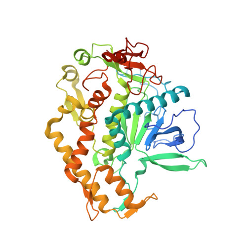
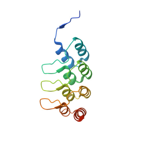
Crystal structure of the LC/A1-DARPin18 complex


Experimental Data Snapshot

  • Method: X-RAY DIFFRACTION
  • Resolution: 2.70 Å
  • R-Value Free: 
    0.306 (Depositor), 0.305 (DCC) 
  • R-Value Work: 
    0.262 (Depositor), 0.260 (DCC) 
  • R-Value Observed: 
    0.264 (Depositor) 

Starting Model: experimental
View more details

wwPDB Validation 3D Report Full Report

Validation slider image for 8HKH

This is version 1.1 of the entry. See complete history. 

Literature

A DARPin promotes faster onset of botulinum neurotoxin A1 action.

Leka, O., Wu, Y., Zanetti, G., Furler, S., Reinberg, T., Marinho, J., Schaefer, J.V., Pluckthun, A., Li, X., Pirazzini, M., Kammerer, R.A.

(2023) Nat Commun 14: 8317-8317

  • DOI: https://doi.org/10.1038/s41467-023-44102-4
  • Primary Citation Related Structures: 
    8HKH

  • PubMed Abstract: 

    In this study, we characterize Designed Ankyrin Repeat Proteins (DARPins) as investigative tools to probe botulinum neurotoxin A1 (BoNT/A1) structure and function. We identify DARPin-F5 that completely blocks SNAP25 substrate cleavage by BoNT/A1 in vitro. X-ray crystallography reveals that DARPin-F5 inhibits BoNT/A1 activity by interacting with a substrate-binding region between the α- and β-exosite. This DARPin does not block substrate cleavage of BoNT/A3, indicating that DARPin-F5 is a subtype-specific inhibitor. BoNT/A1 Glu-171 plays a critical role in the interaction with DARPin-F5 and its mutation to Asp, the residue found in BoNT/A3, results in a loss of inhibition of substrate cleavage. In contrast to the in vitro results, DARPin-F5 promotes faster substrate cleavage of BoNT/A1 in primary neurons and muscle tissue by increasing toxin translocation. Our findings could have important implications for the application of BoNT/A1 in therapeutic areas requiring faster onset of toxin action combined with long persistence.


  • Organizational Affiliation: 
    • Laboratory of Biomolecular Research, Division of Biology, Paul Scherrer Institut, 5232, Villigen PSI, Switzerland.

Macromolecule Content 

  • Total Structure Weight: 132.72 kDa 
  • Atom Count: 8,868 
  • Modeled Residue Count: 1,108 
  • Deposited Residue Count: 1,170 
  • Unique protein chains: 2

Macromolecules

Find similar proteins by:|  3D Structure
Entity ID: 1
MoleculeChains  Sequence LengthOrganismDetailsImage
BoNT/A
A, B
436Clostridium botulinumMutation(s): 0 
Gene Names: a
UniProt
Find proteins for P0DPI1 (Clostridium botulinum (strain Hall / ATCC 3502 / NCTC 13319 / Type A))
Explore P0DPI1 
Go to UniProtKB:  P0DPI1
Entity Groups
Sequence Clusters30% Identity50% Identity70% Identity90% Identity95% Identity100% Identity
UniProt GroupP0DPI1
Sequence Annotations
Expand
Reference Sequence
Find similar proteins by:|  3D Structure
Entity ID: 2
MoleculeChains  Sequence LengthOrganismDetailsImage
Designed ankyrin repeat protein 18
C, D
149synthetic constructMutation(s): 0 
Entity Groups
Sequence Clusters30% Identity50% Identity70% Identity90% Identity95% Identity100% Identity
Sequence Annotations
Expand
Reference Sequence

Experimental Data & Validation

Experimental Data

  • Method: X-RAY DIFFRACTION
  • Resolution: 2.70 Å
  • R-Value Free:  0.306 (Depositor), 0.305 (DCC) 
  • R-Value Work:  0.262 (Depositor), 0.260 (DCC) 
  • R-Value Observed: 0.264 (Depositor) 
Space Group: P 1
Unit Cell:
Length ( Å )Angle ( ˚ )
a = 59.56α = 74.98
b = 59.63β = 89.9
c = 102.18γ = 73.15
Software Package:
Software NamePurpose
PHENIXrefinement
PDB_EXTRACTdata extraction
XDSdata reduction
XSCALEdata scaling
PHASERphasing

Structure Validation

View Full Validation Report


Entry History 

& Funding Information

Deposition Data


Funding OrganizationLocationGrant Number
Swiss National Science FoundationSwitzerland31003A_163449
Swiss National Science FoundationSwitzerland31003A_170028

Revision History  (Full details and data files)

  • Version 1.0: 2023-11-29
    Type: Initial release
  • Version 1.1: 2025-01-01
    Changes: Database references, Structure summary