


The crystal structure of Hu protein in Staphylococcus aureus
Liu, Y.H., Li, Y., Liu, B.To be published.
Experimental Data Snapshot
Starting Model: in silico
View more details
wwPDB Validation 3D Report Full Report

Entity ID: 1 | |||||
|---|---|---|---|---|---|
| Molecule | Chains | Sequence Length | Organism | Details | Image |
| DNA-binding protein HU | A, B [auth C], C [auth B], D | 90 | Staphylococcus aureus subsp. aureus Mu50 | Mutation(s): 0 Gene Names: hup, SAV1473 | 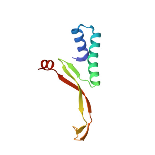 |
UniProt | |||||
Entity Groups | |||||
| Sequence Clusters | 30% Identity50% Identity70% Identity90% Identity95% Identity100% Identity | ||||
| UniProt Group | Q99U17 | ||||
Sequence AnnotationsExpand | |||||
Reference Sequence | |||||
| Length ( Å ) | Angle ( ˚ ) |
|---|---|
| a = 41.427 | α = 90 |
| b = 82.369 | β = 93.157 |
| c = 61.633 | γ = 90 |
| Software Name | Purpose |
|---|---|
| PHENIX | refinement |
| XDS | data reduction |
| Aimless | data scaling |
| PHASER | phasing |
| Funding Organization | Location | Grant Number |
|---|---|---|
| Chinese Academy of Sciences | China | 2020M682420 |


