Biochemical and structural characterization reveals Rv3400 codes for beta-phosphoglucomutase in Mycobacterium tuberculosis.
Singh, L., Karthikeyan, S., Thakur, K.G.(2024) Protein Sci 33: e4943-e4943
- PubMed: 38501428 Search on PubMedSearch on PubMed Central
- DOI: https://doi.org/10.1002/pro.4943
- Primary Citation Related Structures:
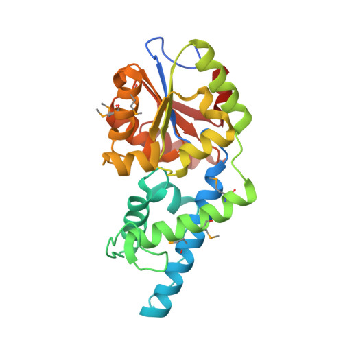
8H5S - PubMed Abstract:
Mycobacterium tuberculosis (Mtb) adapt to various host environments and utilize a variety of sugars and lipids as carbon sources. Among these sugars, maltose and trehalose, also play crucial role in bacterial physiology and virulence. However, some key enzymes involved in trehalose and maltose metabolism in Mtb are not yet known. Here we structurally and functionally characterized a conserved hypothetical gene Rv3400. We determined the crystal structure of Rv3400 at 1.7 Å resolution. The crystal structure revealed that Rv3400 adopts Rossmann fold and shares high structural similarity with haloacid dehalogenase family of proteins. Our comparative structural analysis suggested that Rv3400 could perform either phosphatase or pyrophosphatase or β-phosphoglucomutase (β-PGM) activity. Using biochemical studies, we further confirmed that Rv3400 performs β-PGM activity and hence, Rv3400 encodes for β-PGM in Mtb. Our data also confirm that Mtb β-PGM is a metal dependent enzyme having broad specificity for divalent metal ions. β-PGM converts β-D-glucose-1-phosphate to β-D-glucose-6-phosphate which is required for the generation of ATP and NADPH through glycolysis and pentose phosphate pathway, respectively. Using site directed mutagenesis followed by biochemical studies, we show that two Asp residues in the highly conserved DxD motif, D29 and D31, are crucial for enzyme activity. While D29A, D31A, D29E, D31E and D29N mutants lost complete activity, D31N mutant retained about 30% activity. This study further helps in understanding the role of β-PGM in the physiology of Mtb.
- Division of Protein Science and Engineering, Council of Scientific and Industrial Research-Institute of Microbial Technology (CSIR-IMTECH), Chandigarh, India.
Organizational Affiliation: