8H4O | pdb_00008h4o

Crystal Structure of nucleotide-free Irgb6_T95D mutant


Experimental Data Snapshot

  • Method: X-RAY DIFFRACTION
  • Resolution: 2.05 Å
  • R-Value Free: 
    0.283 (Depositor), 0.280 (DCC) 
  • R-Value Work: 
    0.226 (Depositor), 0.220 (DCC) 
  • R-Value Observed: 
    0.228 (Depositor) 

wwPDB Validation   3D Report Full Report


This is version 1.1 of the entry. See complete history


Literature

Structural basis of Irgb6 inactivation by Toxoplasma gondii through the phosphorylation of switch I.

Okuma, H.Saijo-Hamano, Y.Yamada, H.Sherif, A.A.Hashizaki, E.Sakai, N.Kato, T.Imasaki, T.Kikkawa, S.Nitta, E.Sasai, M.Abe, T.Sugihara, F.Maniwa, Y.Kosako, H.Takei, K.Standley, D.M.Yamamoto, M.Nitta, R.

(2024) Genes Cells 29: 17-38

  • DOI: https://doi.org/10.1111/gtc.13080
  • Primary Citation of Related Structures:  
    8H4O

  • PubMed Abstract: 

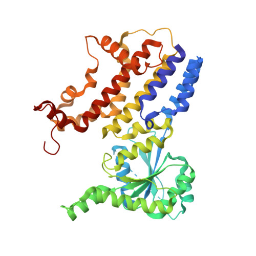
    Irgb6 is a priming immune-related GTPase (IRG) that counteracts Toxoplasma gondii. It is known to be recruited to the low virulent type II T. gondii parasitophorous vacuole (PV), initiating cell-autonomous immunity. However, the molecular mechanism by which immunity-related GTPases become inactivated after the parasite infection remains obscure. Here, we found that Thr95 of Irgb6 is prominently phosphorylated in response to low virulent type II T. gondii infection. We observed that a phosphomimetic T95D mutation in Irgb6 impaired its localization to the PV and exhibited reduced GTPase activity in vitro. Structural analysis unveiled an atypical conformation of nucleotide-free Irgb6-T95D, resulting from a conformational change in the G-domain that allosterically modified the PV membrane-binding interface. In silico docking corroborated the disruption of the physiological membrane binding site. These findings provide novel insights into a T. gondii-induced allosteric inactivation mechanism of Irgb6.


  • Organizational Affiliation
    • Division of Structural Medicine and Anatomy, Department of Physiology and Cell Biology, Kobe University Graduate School of Medicine, Kobe, Japan.

Macromolecules
Find similar proteins by:  (by identity cutoff)  |  3D Structure
Entity ID: 1
MoleculeChains Sequence LengthOrganismDetailsImage
T-cell-specific guanine nucleotide triphosphate-binding protein 2
A, B
417Mus musculusMutation(s): 1 
Gene Names: Tgtp2Ifggb6Irgb6Mg21Tgtp
EC: 3.6.5
UniProt
Find proteins for Q3T9E4 (Mus musculus)
Explore Q3T9E4 
Go to UniProtKB:  Q3T9E4
Entity Groups  
Sequence Clusters30% Identity50% Identity70% Identity90% Identity95% Identity100% Identity
UniProt GroupQ3T9E4
Sequence Annotations
Expand
  • Reference Sequence
Experimental Data & Validation

Experimental Data

  • Method: X-RAY DIFFRACTION
  • Resolution: 2.05 Å
  • R-Value Free:  0.283 (Depositor), 0.280 (DCC) 
  • R-Value Work:  0.226 (Depositor), 0.220 (DCC) 
  • R-Value Observed: 0.228 (Depositor) 
Space Group: P 21 21 21
Unit Cell:
Length ( Å )Angle ( ˚ )
a = 71.532α = 90
b = 80.613β = 90
c = 147.649γ = 90
Software Package:
Software NamePurpose
PHENIXrefinement
XDSdata reduction
XDSdata scaling
PHENIXphasing

Structure Validation

View Full Validation Report


Entry History & Funding Information

Deposition Data


Funding OrganizationLocationGrant Number
Japan Agency for Medical Research and Development (AMED)JapanJP21gm0810013
Japan Society for the Promotion of Science (JSPS)Japan21K06988
Japan Society for the Promotion of Science (JSPS)Japan21H05254
Japan Society for the Promotion of Science (JSPS)Japan21K19352
Japan Society for the Promotion of Science (JSPS)Japan22H02795
Japan Science and TechnologyJapanJPMJMS2024

Revision History  (Full details and data files)

  • Version 1.0: 2023-10-18
    Type: Initial release
  • Version 1.1: 2024-07-03
    Changes: Database references