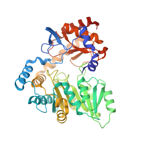
Structural insights into the substrate recognition of serine palmitoyltransferase from Sphingobacterium multivorum.
Ikushiro, H., Murakami, T., Takahashi, A., Katayama, A., Sawai, T., Goto, H., Koolath, S., Murai, Y., Monde, K., Miyahara, I., Kamiya, N., Yano, T.(2023) J Biological Chem 299: 104684-104684
- PubMed: 37030501 Search on PubMedSearch on PubMed Central
- DOI: https://doi.org/10.1016/j.jbc.2023.104684
- Primary Citation Related Structures:
8H1Q, 8H1W, 8H1Y, 8H20, 8H21, 8H29 - PubMed Abstract:
Serine palmitoyltransferase (SPT) is a key enzyme of sphingolipid biosynthesis, which catalyzes the pyridoxal-5'-phosphate-dependent decarboxylative condensation reaction of l-serine (l-Ser) and palmitoyl-CoA (PalCoA) to form 3-ketodihydrosphingosine called long chain base (LCB). SPT is also able to metabolize l-alanine (l-Ala) and glycine (Gly), albeit with much lower efficiency. Human SPT is a membrane-bound large protein complex containing SPTLC1/SPTLC2 heterodimer as the core subunits, and it is known that mutations of the SPTLC1/SPTLC2 genes increase the formation of deoxy-type of LCBs derived from l-Ala and Gly to cause some neurodegenerative diseases. In order to study the substrate recognition of SPT, we examined the reactivity of Sphingobacterium multivorum SPT on various amino acids in the presence of PalCoA. The S. multivorum SPT could convert not only l-Ala and Gly but also l-homoserine, in addition to l-Ser, into the corresponding LCBs. Furthermore, we obtained high-quality crystals of the ligand-free form and the binary complexes with a series of amino acids, including a nonproductive amino acid, l-threonine, and determined the structures at 1.40 to 1.55 Å resolutions. The S. multivorum SPT accommodated various amino acid substrates through subtle rearrangements of the active-site amino acid residues and water molecules. It was also suggested that non-active-site residues mutated in the human SPT genes might indirectly influence the substrate specificity by affecting the hydrogen-bonding networks involving the bound substrate, water molecules, and amino acid residues in the active site of this enzyme. Collectively, our results highlight SPT structural features affecting substrate specificity for this stage of sphingolipid biosynthesis.
- Department of Biochemistry, Faculty of Medicine, Osaka Medical and Pharmaceutical University, Takatsuki, Osaka, Japan. Electronic address: hiroko.ikushiro@ompu.ac.jp.
Organizational Affiliation: