8H1J

Cryo-EM structure of the TnpB-omegaRNA-target DNA ternary complex


Experimental Data Snapshot

  • Method: ELECTRON MICROSCOPY
  • Resolution: 3.10 Å
  • Aggregation State: PARTICLE 
  • Reconstruction Method: SINGLE PARTICLE 

wwPDB Validation   3D Report Full Report


This is version 1.2 of the entry. See complete history


Literature

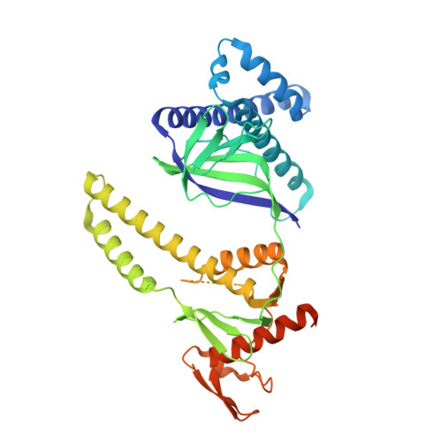
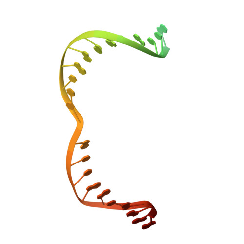
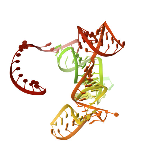
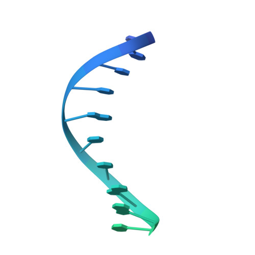
Cryo-EM structure of the transposon-associated TnpB enzyme.

Nakagawa, R.Hirano, H.Omura, S.N.Nety, S.Kannan, S.Altae-Tran, H.Yao, X.Sakaguchi, Y.Ohira, T.Wu, W.Y.Nakayama, H.Shuto, Y.Tanaka, T.Sano, F.K.Kusakizako, T.Kise, Y.Itoh, Y.Dohmae, N.van der Oost, J.Suzuki, T.Zhang, F.Nureki, O.

(2023) Nature 616: 390-397

  • DOI: https://doi.org/10.1038/s41586-023-05933-9
  • Primary Citation of Related Structures:  
    8H1J

  • PubMed Abstract: 

    The class 2 type V CRISPR effector Cas12 is thought to have evolved from the IS200/IS605 superfamily of transposon-associated TnpB proteins 1 . Recent studies have identified TnpB proteins as miniature RNA-guided DNA endonucleases 2,3 . TnpB associates with a single, long RNA (ωRNA) and cleaves double-stranded DNA targets complementary to the ωRNA guide. However, the RNA-guided DNA cleavage mechanism of TnpB and its evolutionary relationship with Cas12 enzymes remain unknown. Here we report the cryo-electron microscopy (cryo-EM) structure of Deinococcus radiodurans ISDra2 TnpB in complex with its cognate ωRNA and target DNA. In the structure, the ωRNA adopts an unexpected architecture and forms a pseudoknot, which is conserved among all guide RNAs of Cas12 enzymes. Furthermore, the structure, along with our functional analysis, reveals how the compact TnpB recognizes the ωRNA and cleaves target DNA complementary to the guide. A structural comparison of TnpB with Cas12 enzymes suggests that CRISPR-Cas12 effectors acquired an ability to recognize the protospacer-adjacent motif-distal end of the guide RNA-target DNA heteroduplex, by either asymmetric dimer formation or diverse REC2 insertions, enabling engagement in CRISPR-Cas adaptive immunity. Collectively, our findings provide mechanistic insights into TnpB function and advance our understanding of the evolution from transposon-encoded TnpB proteins to CRISPR-Cas12 effectors.


  • Organizational Affiliation

    Department of Biological Sciences, Graduate School of Science, The University of Tokyo, Tokyo, Japan.


Macromolecules

Find similar proteins by:  (by identity cutoff)  |  3D Structure
Entity ID: 1
MoleculeChains Sequence LengthOrganismDetailsImage
RNA-guided DNA endonuclease TnpB410Deinococcus radiodurans R1 = ATCC 13939 = DSM 20539Mutation(s): 0 
Gene Names: tnpB
EC: 3.1.21
UniProt
Find proteins for Q7DF80 (Deinococcus radiodurans (strain ATCC 13939 / DSM 20539 / JCM 16871 / CCUG 27074 / LMG 4051 / NBRC 15346 / NCIMB 9279 / VKM B-1422 / R1))
Explore Q7DF80 
Go to UniProtKB:  Q7DF80
Entity Groups  
Sequence Clusters30% Identity50% Identity70% Identity90% Identity95% Identity100% Identity
UniProt GroupQ7DF80
Sequence Annotations
Expand
  • Reference Sequence
Find similar nucleic acids by:  (by identity cutoff)  |  3D Structure
Entity ID: 2
MoleculeChains LengthOrganismImage
Target strandB [auth C]35Deinococcus radiodurans R1 = ATCC 13939 = DSM 20539
Sequence Annotations
Expand
  • Reference Sequence
Find similar nucleic acids by:  (by identity cutoff)  |  3D Structure
Entity ID: 3
MoleculeChains LengthOrganismImage
omegaRNA (130-MER)C [auth B]247Deinococcus radiodurans R1 = ATCC 13939 = DSM 20539
Sequence Annotations
Expand
  • Reference Sequence
Find similar nucleic acids by:  (by identity cutoff)  |  3D Structure
Entity ID: 4
MoleculeChains LengthOrganismImage
Non-target strand35Deinococcus radiodurans R1 = ATCC 13939 = DSM 20539
Sequence Annotations
Expand
  • Reference Sequence
Small Molecules
Ligands 1 Unique
IDChains Name / Formula / InChI Key2D Diagram3D Interactions
ZN (Subject of Investigation/LOI)
Query on ZN

Download Ideal Coordinates CCD File 
E [auth A]ZINC ION
Zn
PTFCDOFLOPIGGS-UHFFFAOYSA-N
Experimental Data & Validation

Experimental Data

  • Method: ELECTRON MICROSCOPY
  • Resolution: 3.10 Å
  • Aggregation State: PARTICLE 
  • Reconstruction Method: SINGLE PARTICLE 

Structure Validation

View Full Validation Report


Entry History & Funding Information

Deposition Data


Funding OrganizationLocationGrant Number
Japan Agency for Medical Research and Development (AMED)JapanJP22ama121012

Revision History  (Full details and data files)

  • Version 1.0: 2023-04-12
    Type: Initial release
  • Version 1.1: 2023-04-19
    Changes: Database references
  • Version 1.2: 2023-04-26
    Changes: Database references