Domain swapping of the C-terminal helix promotes the dimerization of a novel ribonuclease protein from Mycobacterium tuberculosis.
Kim, D.H., Na, Y., Chang, H., Boo, J.H., Kang, S.M., Jin, C., Kang, S.J., Lee, S.Y., Lee, B.J.(2023) Protein Sci 32: e4644-e4644
- PubMed: 37070717 Search on PubMedSearch on PubMed Central
- DOI: https://doi.org/10.1002/pro.4644
- Primary Citation Related Structures:
8H0H, 8H2D - PubMed Abstract:
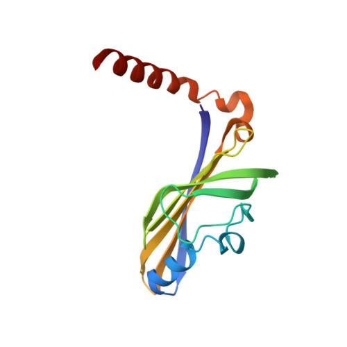
Polyketide metabolism-associated proteins in Mycobacterium tuberculosis play an essential role in the survival of the bacterium, which makes them potential drug targets for the treatment of tuberculosis (TB). The novel ribonuclease protein Rv1546 is predicted to be a member of the steroidogenic acute regulatory protein-related lipid-transfer (START) domain superfamily, which comprises bacterial polyketide aromatase/cyclases (ARO/CYCs). Here, we determined the crystal structure of Rv1546 in a V-shaped dimer. The Rv1546 monomer consists of four α-helices and seven antiparallel β-strands. Interestingly, in the dimeric state, Rv1546 forms a helix-grip fold, which is present in START domain proteins, via three-dimensional domain swapping. Structural analysis revealed that the conformational change of the C-terminal α-helix of Rv1546 might contribute to the unique dimer structure. Site-directed mutagenesis followed by in vitro ribonuclease activity assays was performed to identify catalytic sites of the protein. This experiment suggested that surface residues R63, K84, K88, and R113 are important in the ribonuclease function of Rv1546. In summary, this study presents the structural and functional characterization of Rv1546 and supplies new perspectives for exploiting Rv1546 as a novel drug target for TB treatment.
- Jeju Research Institute of Pharmaceutical Sciences, College of Pharmacy, Jeju National University, Jeju, Korea.
Organizational Affiliation: