8GW8

the human PTH1 receptor bound to an intracellular biased agonist


Experimental Data Snapshot

  • Method: ELECTRON MICROSCOPY
  • Resolution: 2.90 Å
  • Aggregation State: PARTICLE 
  • Reconstruction Method: SINGLE PARTICLE 

wwPDB Validation   3D Report Full Report


This is version 1.3 of the entry. See complete history


Literature

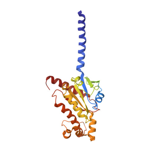
Class B1 GPCR activation by an intracellular agonist.

Kobayashi, K.Kawakami, K.Kusakizako, T.Tomita, A.Nishimura, M.Sawada, K.Okamoto, H.H.Hiratsuka, S.Nakamura, G.Kuwabara, R.Noda, H.Muramatsu, H.Shimizu, M.Taguchi, T.Inoue, A.Murata, T.Nureki, O.

(2023) Nature 618: 1085-1093

  • DOI: https://doi.org/10.1038/s41586-023-06169-3
  • Primary Citation of Related Structures:  
    8GW8

  • PubMed Abstract: 

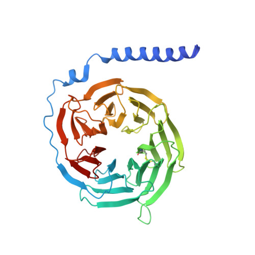
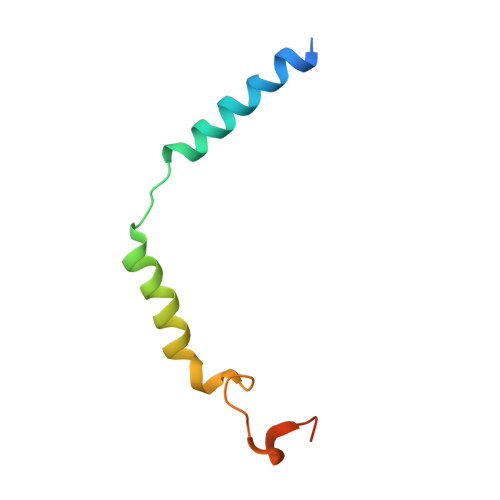
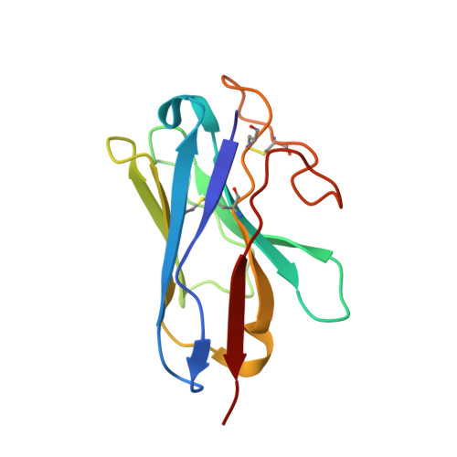
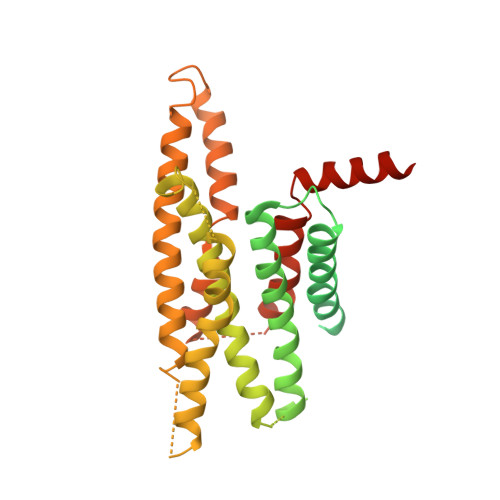
    G protein-coupled receptors (GPCRs) generally accommodate specific ligands in the orthosteric-binding pockets. Ligand binding triggers a receptor allosteric conformational change that leads to the activation of intracellular transducers, G proteins and β-arrestins. Because these signals often induce adverse effects, the selective activation mechanism for each transducer must be elucidated. Thus, many orthosteric-biased agonists have been developed, and intracellular-biased agonists have recently attracted broad interest. These agonists bind within the receptor intracellular cavity and preferentially tune the specific signalling pathway over other signalling pathways, without allosteric rearrangement of the receptor from the extracellular side 1-3 . However, only antagonist-bound structures are currently available 1,4-6 , and there is no evidence to support that biased agonist binding occurs within the intracellular cavity. This limits the comprehension of intracellular-biased agonism and potential drug development. Here we report the cryogenic electron microscopy structure of a complex of G s and the human parathyroid hormone type 1 receptor (PTH1R) bound to a PTH1R agonist, PCO371. PCO371 binds within an intracellular pocket of PTH1R and directly interacts with G s . The PCO371-binding mode rearranges the intracellular region towards the active conformation without extracellularly induced allosteric signal propagation. PCO371 stabilizes the significantly outward-bent conformation of transmembrane helix 6, which facilitates binding to G proteins rather than β-arrestins. Furthermore, PCO371 binds within the highly conserved intracellular pocket, activating 7 out of the 15 class B1 GPCRs. Our study identifies a new and conserved intracellular agonist-binding pocket and provides evidence of a biased signalling mechanism that targets the receptor-transducer interface.


  • Organizational Affiliation

    Department of Biological Sciences, Graduate School of Science, The University of Tokyo, Tokyo, Japan.


Macromolecules
Find similar proteins by:  (by identity cutoff)  |  3D Structure
Entity ID: 1
MoleculeChains Sequence LengthOrganismDetailsImage
Isoform Gnas-2 of Guanine nucleotide-binding protein G(s) subunit alpha isoforms short359Homo sapiensMutation(s): 6 
Gene Names: GNASGNAS1GSP
Membrane Entity: Yes 
UniProt & NIH Common Fund Data Resources
Find proteins for P63092 (Homo sapiens)
Explore P63092 
Go to UniProtKB:  P63092
PHAROS:  P63092
GTEx:  ENSG00000087460 
Entity Groups  
Sequence Clusters30% Identity50% Identity70% Identity90% Identity95% Identity100% Identity
UniProt GroupP63092
Sequence Annotations
Expand
  • Reference Sequence
Find similar proteins by:  (by identity cutoff)  |  3D Structure
Entity ID: 2
MoleculeChains Sequence LengthOrganismDetailsImage
Guanine nucleotide-binding protein G(I)/G(S)/G(T) subunit beta-1351Rattus norvegicusMutation(s): 0 
Gene Names: Gnb1
UniProt
Find proteins for P54311 (Rattus norvegicus)
Explore P54311 
Go to UniProtKB:  P54311
Entity Groups  
Sequence Clusters30% Identity50% Identity70% Identity90% Identity95% Identity100% Identity
UniProt GroupP54311
Sequence Annotations
Expand
  • Reference Sequence
Find similar proteins by:  (by identity cutoff)  |  3D Structure
Entity ID: 3
MoleculeChains Sequence LengthOrganismDetailsImage
Guanine nucleotide-binding protein G(I)/G(S)/G(O) subunit gamma-2C [auth G]68Bos taurusMutation(s): 0 
Gene Names: GNG2
Membrane Entity: Yes 
UniProt
Find proteins for P63212 (Bos taurus)
Explore P63212 
Go to UniProtKB:  P63212
Entity Groups  
Sequence Clusters30% Identity50% Identity70% Identity90% Identity95% Identity100% Identity
UniProt GroupP63212
Sequence Annotations
Expand
  • Reference Sequence
Find similar proteins by:  (by identity cutoff)  |  3D Structure
Entity ID: 4
MoleculeChains Sequence LengthOrganismDetailsImage
Nanobody-35D [auth N]128unidentifiedMutation(s): 0 
Entity Groups  
Sequence Clusters30% Identity50% Identity70% Identity90% Identity95% Identity100% Identity
Sequence Annotations
Expand
  • Reference Sequence
Find similar proteins by:  (by identity cutoff)  |  3D Structure
Entity ID: 5
MoleculeChains Sequence LengthOrganismDetailsImage
Parathyroid hormone/parathyroid hormone-related peptide receptorE [auth R]473Homo sapiensMutation(s): 0 
Gene Names: PTH1RPTHRPTHR1
Membrane Entity: Yes 
UniProt & NIH Common Fund Data Resources
Find proteins for Q03431 (Homo sapiens)
Explore Q03431 
Go to UniProtKB:  Q03431
PHAROS:  Q03431
GTEx:  ENSG00000160801 
Entity Groups  
Sequence Clusters30% Identity50% Identity70% Identity90% Identity95% Identity100% Identity
UniProt GroupQ03431
Sequence Annotations
Expand
  • Reference Sequence
Small Molecules
Ligands 1 Unique
IDChains Name / Formula / InChI Key2D Diagram3D Interactions
KHF
Query on KHF

Download Ideal Coordinates CCD File 
F [auth R]PCO-371
C29 H32 F3 N5 O6 S
LDZJFVOUPUFOHX-UHFFFAOYSA-N
Experimental Data & Validation

Experimental Data

  • Method: ELECTRON MICROSCOPY
  • Resolution: 2.90 Å
  • Aggregation State: PARTICLE 
  • Reconstruction Method: SINGLE PARTICLE 
EM Software:
TaskSoftware PackageVersion
RECONSTRUCTIONRELION3.1

Structure Validation

View Full Validation Report


Entry History & Funding Information

Deposition Data


Funding OrganizationLocationGrant Number
Japan Society for the Promotion of Science (JSPS)Japan--

Revision History  (Full details and data files)

  • Version 1.0: 2023-06-14
    Type: Initial release
  • Version 1.1: 2023-06-21
    Changes: Database references
  • Version 1.2: 2023-07-12
    Changes: Database references, Refinement description
  • Version 1.3: 2023-09-27
    Changes: Data collection, Derived calculations, Structure summary