8GUO

Crystal structure of the nuclease domain of EsaD in complex with EsaG from Staphylococcus aureus


Experimental Data Snapshot

  • Method: X-RAY DIFFRACTION
  • Resolution: 2.59 Å
  • R-Value Free: 0.226 
  • R-Value Work: 0.206 
  • R-Value Observed: 0.208 

wwPDB Validation   3D Report Full Report


This is version 1.1 of the entry. See complete history


Literature

A toxin-deformation dependent inhibition mechanism in the T7SS toxin-antitoxin system of Gram-positive bacteria.

Wang, Y.Zhou, Y.Shi, C.Liu, J.Lv, G.Huang, H.Li, S.Duan, L.Zheng, X.Liu, Y.Zhou, H.Wang, Y.Li, Z.Ding, K.Sun, P.Huang, Y.Lu, X.Zhang, Z.M.

(2022) Nat Commun 13: 6434-6434

  • DOI: https://doi.org/10.1038/s41467-022-34034-w
  • Primary Citation of Related Structures:  
    8GUN, 8GUO, 8GUP

  • PubMed Abstract: 

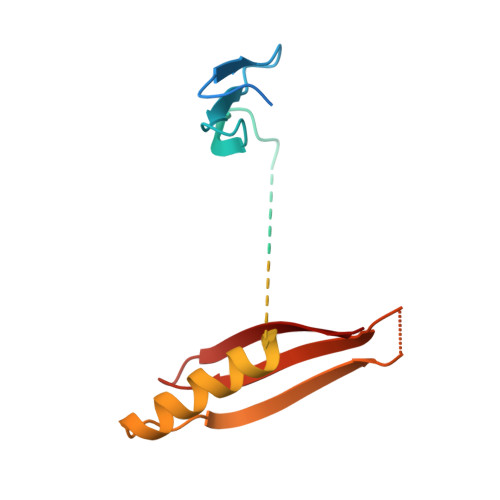
    Toxin EsaD secreted by some S. aureus strains through the type VII secretion system (T7SS) specifically kills those strains lacking the antitoxin EsaG. Here we report the structures of EsaG, the nuclease domain of EsaD and their complex, which together reveal an inhibition mechanism that relies on significant conformational change of the toxin. To inhibit EsaD, EsaG breaks the nuclease domain of EsaD protein into two independent fragments that, in turn, sandwich EsaG. The originally well-folded ββα-metal finger connecting the two fragments is stretched to become a disordered loop, leading to disruption of the catalytic site of EsaD and loss of nuclease activity. This mechanism is distinct from that of the other Type II toxin-antitoxin systems, which utilize an intrinsically disordered region on the antitoxins to cover the active site of the toxins. This study paves the way for developing therapeutic approaches targeting this antagonism.


  • Organizational Affiliation

    International Cooperative Laboratory of Traditional Chinese Medicine Modernization and Innovative Drug Development of Chinese Ministry of Education (MOE), College of Pharmacy, Jinan University, Guangzhou, 510632, China.


Macromolecules
Find similar proteins by:  (by identity cutoff)  |  3D Structure
Entity ID: 1
MoleculeChains Sequence LengthOrganismDetailsImage
Type VII secretion system protein EsaG162Staphylococcus aureus subsp. aureus NCTC 8325Mutation(s): 0 
Gene Names: essGesaGSAOUHSC_00269
UniProt
Find proteins for Q2G178 (Staphylococcus aureus (strain NCTC 8325 / PS 47))
Explore Q2G178 
Go to UniProtKB:  Q2G178
Entity Groups  
Sequence Clusters30% Identity50% Identity70% Identity90% Identity95% Identity100% Identity
UniProt GroupQ2G178
Sequence Annotations
Expand
  • Reference Sequence
Find similar proteins by:  (by identity cutoff)  |  3D Structure
Entity ID: 2
MoleculeChains Sequence LengthOrganismDetailsImage
Type VII secretion system protein EssD164Staphylococcus aureus subsp. aureus NCTC 8325Mutation(s): 0 
Gene Names: essDesaDSAOUHSC_00268
UniProt
Find proteins for Q2G179 (Staphylococcus aureus (strain NCTC 8325 / PS 47))
Explore Q2G179 
Go to UniProtKB:  Q2G179
Entity Groups  
Sequence Clusters30% Identity50% Identity70% Identity90% Identity95% Identity100% Identity
UniProt GroupQ2G179
Sequence Annotations
Expand
  • Reference Sequence
Experimental Data & Validation

Experimental Data

  • Method: X-RAY DIFFRACTION
  • Resolution: 2.59 Å
  • R-Value Free: 0.226 
  • R-Value Work: 0.206 
  • R-Value Observed: 0.208 
  • Space Group: I 4 2 2
Unit Cell:
Length ( Å )Angle ( ˚ )
a = 111.776α = 90
b = 111.776β = 90
c = 171.228γ = 90
Software Package:
Software NamePurpose
PHENIXrefinement
HKL-3000data reduction
HKL-2000data scaling
PHASERphasing
Cootmodel building

Structure Validation

View Full Validation Report


Entry History & Funding Information

Deposition Data


Funding OrganizationLocationGrant Number
Not funded--

Revision History  (Full details and data files)

  • Version 1.0: 2022-11-09
    Type: Initial release
  • Version 1.1: 2024-04-03
    Changes: Data collection, Refinement description