Structural insights of the toxin-antitoxin system VPA0770-VPA0769 in Vibrio parahaemolyticus.
Zhang, Y., Song, X., Chen, C., Liu, L., Xu, Y., Zhang, N., Huang, W., Zheng, J., Yuan, W., Tang, L., Lin, Z.(2023) Int J Biol Macromol 242: 124755-124755
- PubMed: 37164131 Search on PubMed
- DOI: https://doi.org/10.1016/j.ijbiomac.2023.124755
- Primary Citation Related Structures:
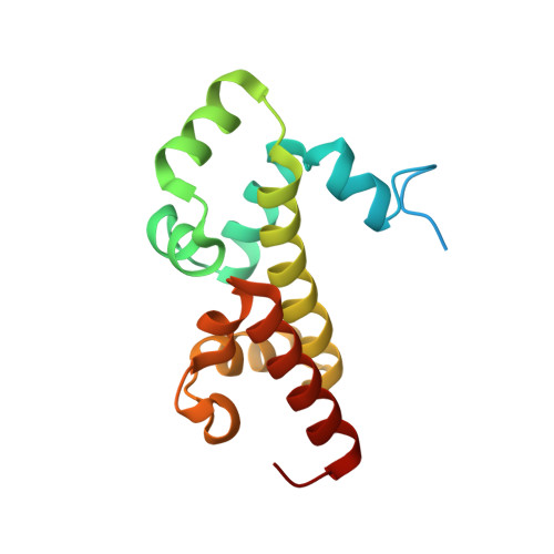
8GUG - PubMed Abstract:
Toxin-antitoxin (TA) systems are involved in both normal bacterial physiology and pathogenicity, including gene regulation, antibiotic resistance, and bacteria persistence under stressful environments. In pathogenic Vibrio parahaemolyticus, however, TA interaction and assembly remain largely unknown. In this work, we identified a new RES-Xre type II TA module, encoded by gene cluster vpa0770-vpa0769 on chromosome II of V. parahaemolyticus. Ectopic expression of the VPA0770 toxin rapidly arrests the growth of E. coli cells, which can be neutralized by co-expression of the VPA0769 antitoxin. To decipher the action mechanism, we determined the crystal structure of the VPA0770-VPA0769 TA complex. VPA0770 and VPA0769 proteins can assemble into two types of large complexes, a W-shaped hetero-hexamer and a donut-like hetero-dodecamer, in a concentration-dependent manner in solution. Disruption of the TA interface results in a loss of the antitoxic phenotype. The toxicity of the VPA0770 toxin, which harbors a NAD + -binding pocket, may be largely ascribed to its highly effective capability to degrade intracellular NAD + . Our study provides a structural basis for a better understanding of diverse molecular mechanisms employed by human pathogens.
- School of Life Sciences, Tianjin University, Tianjin 300073, China.
Organizational Affiliation: