Find PDB structures and Computed Structure Models (CSM) by combining queries from tools in this suite: Attribute Search, Sequence Similarity, Sequence Motif, 3D Similarity, and 3D Motif with 'AND' logic.
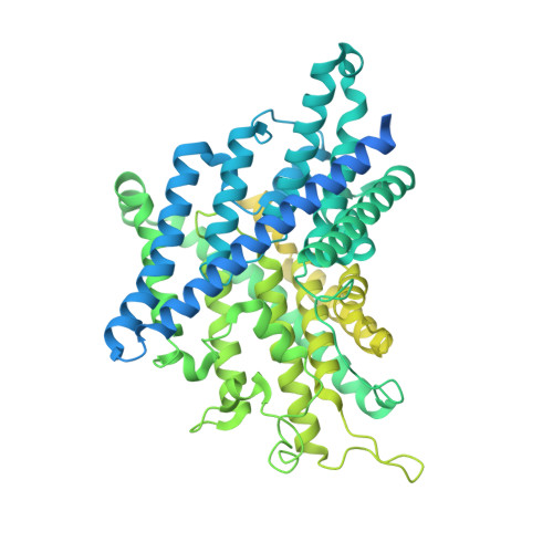
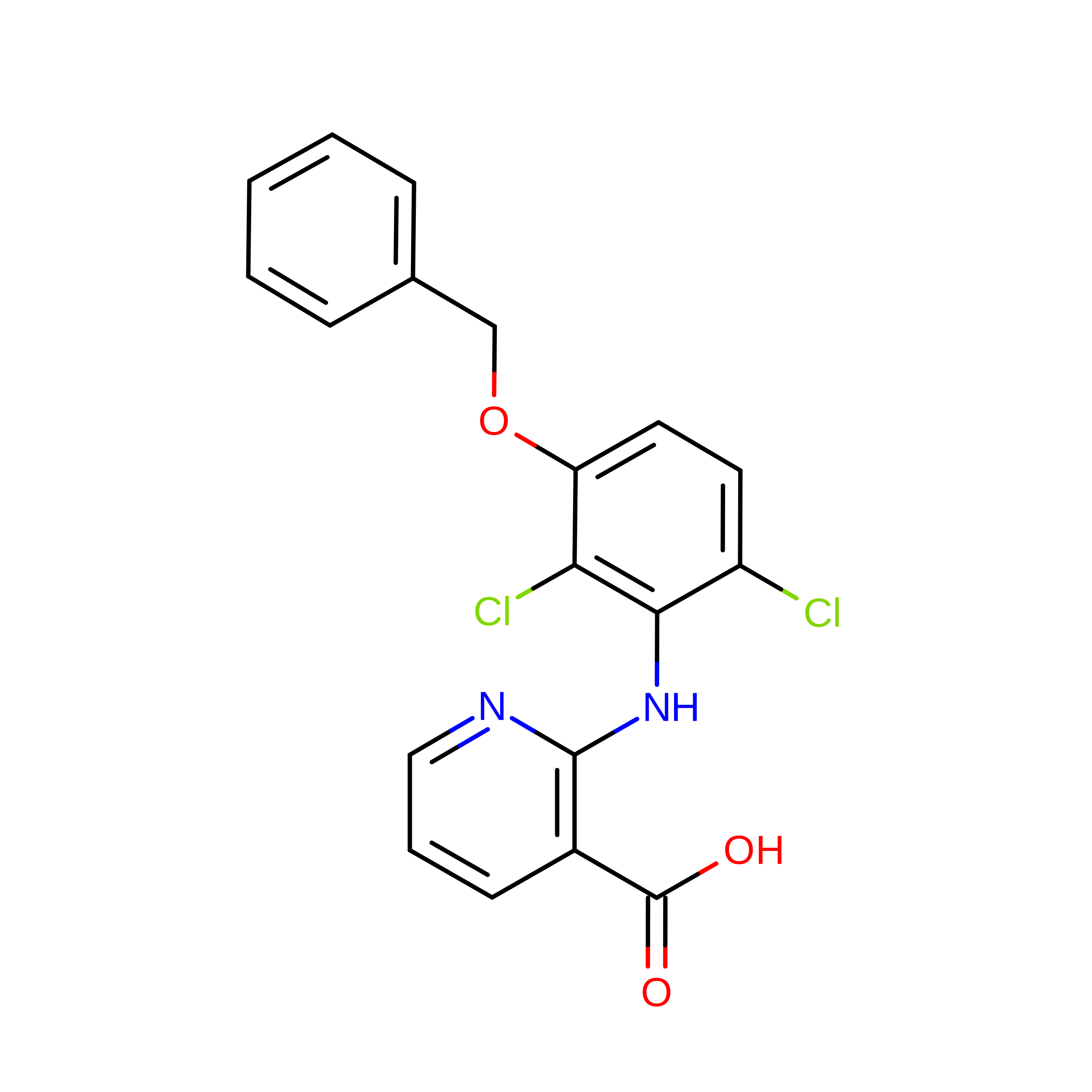
ClC-2 transports chloride ions across plasma membranes and plays critical roles in cellular homeostasis. Its dysfunction is involved in diseases including leukodystrophy and primary aldosteronism. AK-42 was recently reported as a specific inhibitor of ClC-2. However, experimental structures are still missing to decipher its inhibition mechanism. Here, we present cryo-EM structures of apo ClC-2 and its complex with AK-42, both at 3.5 Å resolution. Residues S162, E205 and Y553 are involved in chloride binding and contribute to the ion selectivity. The side-chain of the gating glutamate E205 occupies the putative central chloride-binding site, indicating that our structure represents a closed state. Structural analysis, molecular dynamics and electrophysiological recordings identify key residues to interact with AK-42. Several AK-42 interacting residues are present in ClC-2 but not in other ClCs, providing a possible explanation for AK-42 specificity. Taken together, our results experimentally reveal the potential inhibition mechanism of ClC-2 inhibitor AK-42.
Organizational Affiliation:
Shenzhen Institute of Advanced Technology, Chinese Academy of Sciences, 518055, Shenzhen, China.
Department of Biomedical Engineering, Southern University of Science and Technology, 518055, Shenzhen, China.
School of Life Sciences, Southern University of Science and Technology, 518055, Shenzhen, China.
Shenzhen Key Laboratory of Translational Research for Brain Diseases, The Brain Cognition and Brain Disease Institute, Shenzhen Institute of Advanced Technology, Chinese Academy of Sciences, Shenzhen, China.
Shenzhen-Hong Kong Institute of Brain Science-Shenzhen Fundamental Research Institutions, 518055, Shenzhen, Guangdong, China.
University of Chinese Academy of Sciences, Beijing, China.
School of Life Sciences, The Chinese University of Hong Kong, Shatin, Hong Kong, China.
Interdisciplinary Center for Brain Information, The Brain Cognition and Brain Disease Institute, Shenzhen Institute of Advanced Technology, Chinese Academy of Sciences, 518055, Shenzhen, Guangdong, China.
Faculty of Life and Health Sciences, Shenzhen Institute of Advanced Technology, Chinese Academy of Sciences, 518055, Shenzhen, Guangdong, China.
Department of Child and Adolescent Psychiatry, Shenzhen Kangning Hospital, Shenzhen Mental Health Center, Shenzhen, 518020, China.
Cryo-EM Facility Center, Southern University of Science and Technology, 518055, Shenzhen, Guangdong, China.
Shenzhen Institute of Advanced Technology, Chinese Academy of Sciences, 518055, Shenzhen, China. horst.vogel@siat.ac.cn.
Institut des Sciences et Ingénierie Chimiques (ISIC), Ecole Polytechnique Fédérale de Lausanne (EPFL), Lausanne, Switzerland. horst.vogel@siat.ac.cn.
Shenzhen Key Laboratory of Translational Research for Brain Diseases, The Brain Cognition and Brain Disease Institute, Shenzhen Institute of Advanced Technology, Chinese Academy of Sciences, Shenzhen, China. wangyt@siat.ac.cn.
Faculty of Life and Health Sciences, Shenzhen Institute of Advanced Technology, Chinese Academy of Sciences, 518055, Shenzhen, Guangdong, China. wangyt@siat.ac.cn.
Department of Biomedical Engineering, Southern University of Science and Technology, 518055, Shenzhen, China. wangdp@mail.sustech.edu.cn.
Department of Orthopedics, Shenzhen Intelligent Orthopaedics and Biomedical Innovation Platform, Guangdong Provincial Research Center for Artificial Intelligence and Digital Orthopedic Technology, Shenzhen Second People's Hospital, The First Affiliated Hospital of Shenzhen University, 518000, Shenzhen, China. wangdp@mail.sustech.edu.cn.
School of Life Sciences, Southern University of Science and Technology, 518055, Shenzhen, China. yankg@sustech.edu.cn.
Shenzhen Institute of Advanced Technology, Chinese Academy of Sciences, 518055, Shenzhen, China. zhanghw@sustech.edu.cn.
Department of Biomedical Engineering, Southern University of Science and Technology, 518055, Shenzhen, China. zhanghw@sustech.edu.cn.

 Download Mendeley