Unravelling biochemical and structural features of Bacillus licheniformis GH5 mannanase using site-directed mutagenesis and high-resolution protein crystallography studies.
Briganti, L., Manzine, L.R., de Mello Capetti, C.C., de Araujo, E.A., de Oliveira Arnoldi Pellegrini, V., Guimaraes, F.E.G., de Oliveira Neto, M., Polikarpov, I.(2024) Int J Biol Macromol 274: 133182-133182
- PubMed: 38885857 Search on PubMed
- DOI: https://doi.org/10.1016/j.ijbiomac.2024.133182
- Primary Citation Related Structures:
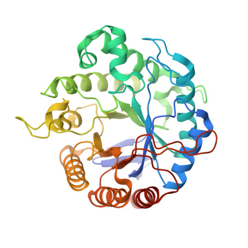
8GF3 - PubMed Abstract:
Glycoside hydrolase family 5 (GH5) encompasses enzymes with several different activities, including endo-1,4-β-mannosidases. These enzymes are involved in mannan degradation, and have a number of biotechnological applications, such as mannooligosaccharide prebiotics production, stain removal and dyes decolorization, to name a few. Despite the importance of GH5 enzymes, only a few members of subfamily 7 were structurally characterized. In the present work, biochemical and structural characterization of Bacillus licheniformis GH5 mannanase, BlMan5_7 were performed and the enzyme cleavage pattern was analyzed, showing that BlMan5_7 requires at least 5 occupied subsites to perform efficient hydrolysis. Additionally, crystallographic structure at 1.3 Å resolution was determined and mannoheptaose (M7) was docked into the active site to investigate the interactions between substrate and enzyme through molecular dynamic (MD) simulations, revealing the existence of a - 4 subsite, which might explain the generation of mannotetraose (M4) as an enzyme product. Biotechnological application of the enzyme in stain removal was investigated, demonstrating that BlMan5_7 addition to washing solution greatly improves mannan-based stain elimination.
- Instituto de Física de São Carlos, Universidade de São Paulo, Avenida Trabalhador São Carlense 400 - Centro, São Carlos, SP 13560-970, Brazil.
Organizational Affiliation: