Structural analysis of the water channel AQP2 by single-particle cryo-EM.
Kamegawa, A., Suzuki, S., Suzuki, H., Nishikawa, K., Numoto, N., Fujiyoshi, Y.(2023) J Struct Biol 215: 107984-107984
- PubMed: 37315821 Search on PubMed
- DOI: https://doi.org/10.1016/j.jsb.2023.107984
- Primary Citation Related Structures:
8GCL - PubMed Abstract:
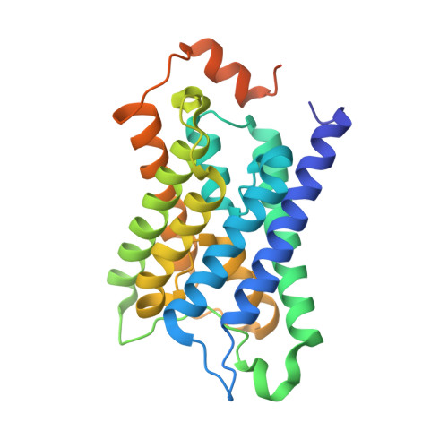
Water channels, which are small membrane proteins almost entirely buried in lipid membranes, are challenging research targets for single-particle cryo-electron microscopy (cryo-EM), a powerful technique routinely used to determine the structures of membrane proteins. Because the single-particle method enables structural analysis of a whole protein with flexible parts that interfere with crystallization, we have focused our efforts on analyzing water channel structures. Here, utilizing this system, we analyzed the structure of full-length aquaporin-2 (AQP2), a primary regulator of vasopressin-dependent reabsorption of water at the renal collecting ducts. The 2.9 Å resolution map revealed a cytoplasmic extension of the cryo-EM density that was presumed to be the highly flexible C-terminus at which the localization of AQP2 is regulated in the renal collecting duct cells. We also observed a continuous density along the common water pathway inside the channel pore and lipid-like molecules at the membrane interface. Observations of these constructions in the AQP2 structure analyzed without any fiducial markers (e.g., a rigidly bound antibody) indicate that single-particle cryo-EM will be useful for investigating water channels in native states as well as in complexes with chemical compounds.
- Cellular and Structural Physiology Laboratory (CeSPL), Tokyo Medical and Dental University, 1-5-45, Yushima, Bunkyo-ku, Tokyo 113-8510, Japan.
Organizational Affiliation: