


Crystal Structure of SETDB1 Tudor domain in complex with UNC6535
Beldar, S., Dong, A., Brown, P.J., Arrowsmith, C.H., Edwards, A.M., Halabelian, L.To be published.
Experimental Data Snapshot
Starting Model: experimental
View more details
Entity ID: 1 | |||||
|---|---|---|---|---|---|
| Molecule | Chains | Sequence Length | Organism | Details | Image |
| Histone-lysine N-methyltransferase SETDB1 | 225 | Homo sapiens | Mutation(s): 0 Gene Names: SETDB1, KIAA0067, KMT1E EC: 2.1.1.43 (PDB Primary Data), 2.1.1.366 (UniProt) | 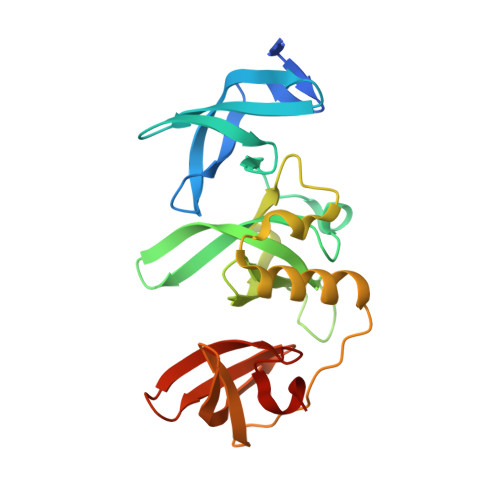 | |
UniProt & NIH Common Fund Data Resources | |||||
PHAROS: Q15047 GTEx: ENSG00000143379 | |||||
Entity Groups | |||||
| Sequence Clusters | 30% Identity50% Identity70% Identity90% Identity95% Identity100% Identity | ||||
| UniProt Group | Q15047 | ||||
Sequence AnnotationsExpand | |||||
Reference Sequence | |||||
| Ligands 2 Unique | |||||
|---|---|---|---|---|---|
| ID | Chains | Name / Formula / InChI Key | 2D Diagram | 3D Interactions | |
| YOH (Subject of Investigation/LOI) Download:Ideal Coordinates CCD File | B [auth A] | N~4~-[6-(dimethylamino)hexyl]-N~2~-[5-(dimethylamino)pentyl]-6,7-dimethoxyquinazoline-2,4-diamine C25 H44 N6 O2 LJBDNFUYQUHYOX-UHFFFAOYSA-N |  | ||
| UNX Download:Ideal Coordinates CCD File | C [auth A] D [auth A] E [auth A] F [auth A] G [auth A] | UNKNOWN ATOM OR ION X |  | ||
| Length ( Å ) | Angle ( ˚ ) |
|---|---|
| a = 54.056 | α = 90 |
| b = 61.997 | β = 90 |
| c = 69.335 | γ = 90 |
| Software Name | Purpose |
|---|---|
| HKL-3000 | data scaling |
| REFMAC | refinement |
| HKL-3000 | data reduction |
| REFMAC | phasing |
| Funding Organization | Location | Grant Number |
|---|---|---|
| Other private | Canada | -- |


