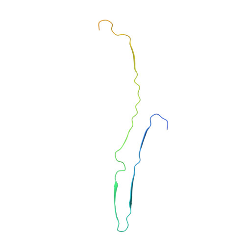
Structure of the nonhelical filament of the Alzheimer's disease tau core.
Duan, P., Dregni, A.J., Mammeri, N.E., Hong, M.(2023) Proc Natl Acad Sci U S A 120: e2310067120-e2310067120
- PubMed: 37878719
- DOI: https://doi.org/10.1073/pnas.2310067120
- Primary Citation of Related Structures:
8G58 - PubMed Abstract:
The microtubule-associated protein tau aggregates into neurofibrillary tangles in Alzheimer's disease (AD). The main type of aggregates, the paired helical filaments (PHF), incorporate about 20% of the full-length protein into the rigid core. Recently, cryo-electron microscopy data showed that a protease-resistant fragment of tau (residues 297-391) self-assembles in vitro in the presence of divalent cations to form twisted filaments whose molecular structure resembles that of AD PHF tau [S. Lövestam et al., Elife 11 , e76494 (2022)]. To investigate whether this tau construct is uniquely predisposed to this morphology and structure, we fibrillized tau (297-391) under the reported conditions and determined its structure using solid-state NMR spectroscopy. Unexpectedly, the protein assembled predominantly into nontwisting ribbons whose rigid core spans residues 305-357. This rigid core forms a β-arch that turns at residues 322 CGS 324 . Two protofilaments stack together via a long interface that stretches from G323 to I354. Together, these two protofilaments form a four-layered β-sheet core whose sidechains are stabilized by numerous polar and hydrophobic interactions. This structure gives insight into the fibril morphologies and molecular conformations that can be adopted by this protease-resistant core of AD tau under different pH and ionic conditions.
- Department of Chemistry, Massachusetts Institute of Technology, Cambridge, MA 02139.
Organizational Affiliation:


