8G2V

Cryo-EM structure of recombinant human LECT2 amyloid fibril core


Experimental Data Snapshot

  • Method: ELECTRON MICROSCOPY
  • Resolution: 2.71 Å
  • Aggregation State: FILAMENT 
  • Reconstruction Method: HELICAL 

wwPDB Validation   3D Report Full Report


This is version 1.2 of the entry. See complete history


Literature

Cryo-EM structure of a human LECT2 amyloid fibril reveals a network of polar ladders at its core.

Richards, L.S.Flores, M.D.Zink, S.Schibrowsky, N.A.Sawaya, M.R.Rodriguez, J.A.

(2023) Structure 31: 1386-1393.e3

  • DOI: https://doi.org/10.1016/j.str.2023.08.007
  • Primary Citation of Related Structures:  
    8G2V

  • PubMed Abstract: 

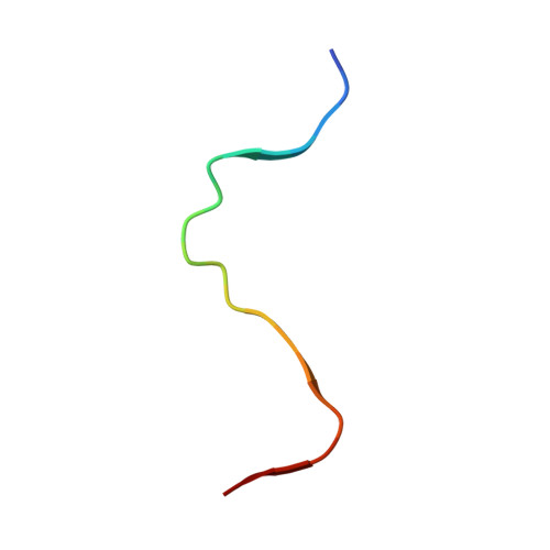
    ALECT2 systemic amyloidosis is associated with deposition of the leukocyte cell-derived chemotaxin-2 (LECT2) protein in the form of fibrils. In ALECT2 amyloidosis, ALECT2 fibrils deposit in the glomerulus, resulting in renal failure. Patients lack effective treatment options outside of renal transplant or dialysis. The structure of globular LECT2 has been determined but structures of ALECT2 amyloid fibrils remain unknown. Using single-particle cryo-EM, we find that recombinant human LECT2 forms robust twisting fibrils with canonical amyloid features. ALECT2 fibrils contain two mating protofilaments spanning residues 55-75 of the LECT2 sequence. The geometry of the ALECT2 fibril displays features in line with other pathogenic amyloids. Its core is tightly packed and stabilized by both hydrophobic contacts and hydrogen-bonded uncharged polar residues. The robustness of ALECT2 fibril cores is illustrated by their resistance to denaturants and proteases. This ALECT2 fibril structure presents a potential new target for treatments against ALECT2 systemic amyloidosis.


  • Organizational Affiliation

    Department of Chemistry and Biochemistry, UCLA-DOE Institute for Genomics and Proteomics, STROBE, NSF Science and Technology Center, University of California, Los Angeles (UCLA), Los Angeles, CA 90095, USA.


Macromolecules

Find similar proteins by:  Sequence   |   3D Structure  

Entity ID: 1
MoleculeChains Sequence LengthOrganismDetailsImage
Leukocyte cell-derived chemotaxin-2
A, B, C, D, E
A, B, C, D, E, F, G, H, I, J
21Homo sapiensMutation(s): 0 
Gene Names: LECT2
UniProt & NIH Common Fund Data Resources
Find proteins for O14960 (Homo sapiens)
Explore O14960 
Go to UniProtKB:  O14960
PHAROS:  O14960
GTEx:  ENSG00000145826 
Entity Groups  
Sequence Clusters30% Identity50% Identity70% Identity90% Identity95% Identity100% Identity
UniProt GroupO14960
Sequence Annotations
Expand
  • Reference Sequence
Experimental Data & Validation

Experimental Data

  • Method: ELECTRON MICROSCOPY
  • Resolution: 2.71 Å
  • Aggregation State: FILAMENT 
  • Reconstruction Method: HELICAL 
EM Software:
TaskSoftware PackageVersion
RECONSTRUCTIONRELION3.1
MODEL REFINEMENTPHENIX

Structure Validation

View Full Validation Report


Entry History & Funding Information

Deposition Data


Funding OrganizationLocationGrant Number
Department of Energy (DOE, United States)United StatesDE-FC02-02ER63421
National Science Foundation (NSF, United States)United StatesDMR-1548924
National Institutes of Health/National Institute of General Medical Sciences (NIH/NIGMS)United StatesGrant R35 GM128867
National Institutes of Health/National Center for Research Resources (NIH/NCRR)United States5T32GM008496
National Institutes of Health/National Center for Research Resources (NIH/NCRR)United StatesU24 GM129541

Revision History  (Full details and data files)

  • Version 1.0: 2023-08-16
    Type: Initial release
  • Version 1.1: 2023-09-13
    Changes: Database references
  • Version 1.2: 2023-11-15
    Changes: Database references