Histidine Protonation and Conformational Switching in Diphtheria Toxin Translocation Domain.
Rodnin, M.V., Vasques-Montes, V., Kyrychenko, A., Oliveira, N.F.B., Kashipathy, M.M., Battaile, K.P., Douglas, J., Lovell, S., Machuqueiro, M., Ladokhin, A.S.(2023) Toxins (Basel) 15
- PubMed: 37505680 Search on PubMedSearch on PubMed Central
- DOI: https://doi.org/10.3390/toxins15070410
- Primary Citation Related Structures:
8G0F, 8G0G - PubMed Abstract:
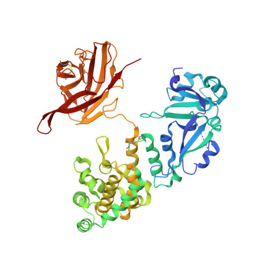
Protonation of key histidine residues has been long implicated in the acid-mediated cellular action of the diphtheria toxin translocation (T-) domain, responsible for the delivery of the catalytic domain into the cell. Here, we use a combination of computational (constant-pH Molecular Dynamics simulations) and experimental (NMR, circular dichroism, and fluorescence spectroscopy along with the X-ray crystallography) approaches to characterize the initial stages of conformational change happening in solution in the wild-type T-domain and in the H223Q/H257Q double mutant. This replacement suppresses the acid-induced transition, resulting in the retention of a more stable protein structure in solutions at pH 5.5 and, consequently, in reduced membrane-disrupting activity. Here, for the first time, we report the pK a values of the histidine residues of the T-domain, measured by NMR-monitored pH titrations. Most peaks in the histidine side chain spectral region are titrated with pK a s ranging from 6.2 to 6.8. However, the two most up-field peaks display little change down to pH 6, which is a limiting pH for this protein in solution at concentrations required for NMR. These peaks are absent in the double mutant, suggesting they belong to H223 and H257. The constant-pH simulations indicate that for the T-domain in solution, the pK a values for histidine residues range from 3.0 to 6.5, with those most difficult to protonate being H251 and H257. Taken together, our experimental and computational data demonstrate that previously suggested cooperative protonation of all six histidines in the T-domain does not occur.
- Department of Biochemistry and Molecular Biology, University of Kansas School of Medicine, Kansas City, KS 66160, USA.
Organizational Affiliation: