Group 3 medulloblastoma transcriptional networks collapse under domain specific EP300/CBP inhibition.
Shendy, N.A.M., Bikowitz, M., Sigua, L.H., Zhang, Y., Mercier, A., Khashana, Y., Nance, S., Liu, Q., Delahunty, I.M., Robinson, S., Goel, V., Rees, M.G., Ronan, M.A., Wang, T., Kocak, M., Roth, J.A., Wang, Y., Freeman, B.B., Orr, B.A., Abraham, B.J., Roussel, M.F., Schonbrunn, E., Qi, J., Durbin, A.D.(2024) Nat Commun 15: 3483-3483
- PubMed: 38664416 Search on PubMedSearch on PubMed Central
- DOI: https://doi.org/10.1038/s41467-024-47102-0
- Primary Citation Related Structures:
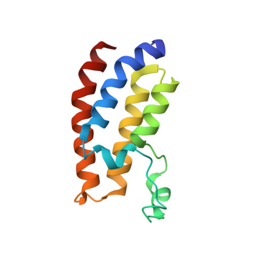
8FV2, 8FVF, 8FVK, 8FVS, 8FXA, 8FXE, 8FXO, 8G6T, 8GA2 - PubMed Abstract:
Chemical discovery efforts commonly target individual protein domains. Many proteins, including the EP300/CBP histone acetyltransferases (HATs), contain several targetable domains. EP300/CBP are critical gene-regulatory targets in cancer, with existing high potency inhibitors of either the catalytic HAT domain or protein-binding bromodomain (BRD). A domain-specific inhibitory approach to multidomain-containing proteins may identify exceptional-responding tumor types, thereby expanding a therapeutic index. Here, we discover that targeting EP300/CBP using the domain-specific inhibitors, A485 (HAT) or CCS1477 (BRD) have different effects in select tumor types. Group 3 medulloblastoma (G3MB) cells are especially sensitive to BRD, compared with HAT inhibition. Structurally, these effects are mediated by the difluorophenyl group in the catalytic core of CCS1477. Mechanistically, bromodomain inhibition causes rapid disruption of genetic dependency networks that are required for G3MB growth. These studies provide a domain-specific structural foundation for drug discovery efforts targeting EP300/CBP and identify a selective role for the EP300/CBP bromodomain in maintaining genetic dependency networks in G3MB.
- Division of Molecular Oncology, Department of Oncology, St. Jude Children's Research Hospital, Memphis, TN, USA.
Organizational Affiliation: