8FNB | pdb_00008fnb

Crystal structure of the C-terminal Fg domain of human TNC with the mutations Y2140H and S2164H

  • Classification: SIGNALING PROTEIN
  • Organism(s): Homo sapiens
  • Expression System: Escherichia coli
  • Mutation(s): Yes 

  • Deposited: 2022-12-27 Released: 2023-07-12 
  • Deposition Author(s): Bouyain, S.
  • Funding Organization(s): National Institutes of Health/National Institute of General Medical Sciences (NIH/NIGMS)

Experimental Data Snapshot

  • Method: X-RAY DIFFRACTION
  • Resolution: 1.80 Å
  • R-Value Free: 
    0.174 (Depositor), 0.191 (DCC) 
  • R-Value Work: 
    0.152 (Depositor) 
  • R-Value Observed: 
    0.153 (Depositor) 

Starting Model: experimental
View more details

wwPDB Validation 3D Report Full Report

Validation slider image for 8FNB

This is version 1.3 of the entry. See complete history

Literature

Protein-protein interactions between tenascin-R and RPTP zeta /phosphacan are critical to maintain the architecture of perineuronal nets.

Sinha, A.Kawakami, J.Cole, K.S.Ladutska, A.Nguyen, M.Y.Zalmai, M.S.Holder, B.L.Broerman, V.M.Matthews, R.T.Bouyain, S.

(2023) J Biological Chem 299: 104952-104952

  • DOI: https://doi.org/10.1016/j.jbc.2023.104952
  • Primary Citation Related Structures: 
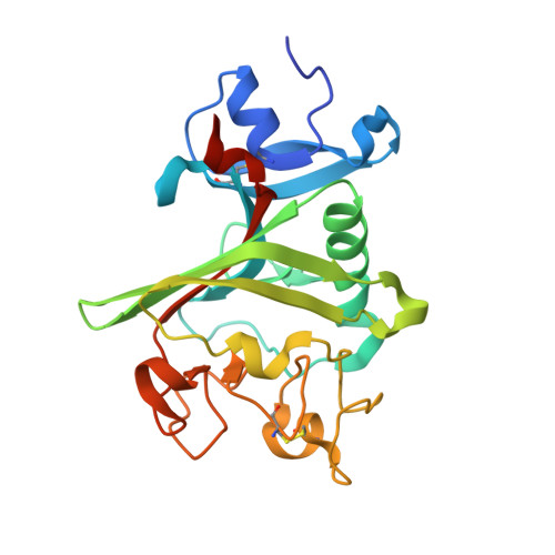
    8FN8, 8FN9, 8FNA, 8FNB

  • PubMed Abstract: 

    Neural plasticity, the ability to alter the structure and function of neural circuits, varies throughout the age of an individual. The end of the hyperplastic period in the central nervous system coincides with the appearance of honeycomb-like structures called perineuronal nets (PNNs) that surround a subset of neurons. PNNs are a condensed form of neural extracellular matrix that include the glycosaminoglycan hyaluronan and extracellular matrix proteins such as aggrecan and tenascin-R (TNR). PNNs are key regulators of developmental neural plasticity and cognitive functions, yet our current understanding of the molecular interactions that help assemble them remains limited. Disruption of Ptprz1, the gene encoding the receptor protein tyrosine phosphatase RPTPζ, altered the appearance of nets from a reticulated structure to puncta on the surface of cortical neuron bodies in adult mice. The structural alterations mirror those found in Tnr -/- mice, and TNR is absent from the net structures that form in dissociated cultures of Ptprz1 -/- cortical neurons. These findings raised the possibility that TNR and RPTPζ cooperate to promote the assembly of PNNs. Here, we show that TNR associates with the RPTPζ ectodomain and provide a structural basis for these interactions. Furthermore, we show that RPTPζ forms an identical complex with tenascin-C, a homolog of TNR that also regulates neural plasticity. Finally, we demonstrate that mutating residues at the RPTPζ-TNR interface impairs the formation of PNNs in dissociated neuronal cultures. Overall, this work sets the stage for analyzing the roles of protein-protein interactions that underpin the formation of nets.


  • Organizational Affiliation
    • Department of Neuroscience and Physiology, State University of New York Upstate Medical University, Syracuse, New York, USA.

Macromolecule Content 

  • Total Structure Weight: 53.1 kDa 
  • Atom Count: 3,999 
  • Modeled Residue Count: 438 
  • Deposited Residue Count: 462 
  • Unique protein chains: 1

Macromolecules

Find similar proteins by:|  3D Structure
Entity ID: 1
MoleculeChains  Sequence LengthOrganismDetailsImage
Tenascin
A, B
231Homo sapiensMutation(s): 2 
Gene Names: TNCHXB
UniProt & NIH Common Fund Data Resources
Find proteins for P24821 (Homo sapiens)
Explore P24821 
Go to UniProtKB:  P24821
PHAROS:  P24821
GTEx:  ENSG00000041982 
Entity Groups
Sequence Clusters30% Identity50% Identity70% Identity90% Identity95% Identity100% Identity
UniProt GroupP24821
Sequence Annotations
Expand
Reference Sequence

Experimental Data & Validation

Experimental Data

  • Method: X-RAY DIFFRACTION
  • Resolution: 1.80 Å
  • R-Value Free:  0.174 (Depositor), 0.191 (DCC) 
  • R-Value Work:  0.152 (Depositor) 
  • R-Value Observed: 0.153 (Depositor) 
Space Group: C 1 2 1
Unit Cell:
Length ( Å )Angle ( ˚ )
a = 106.045α = 90
b = 101.561β = 122.353
c = 56.667γ = 90
Software Package:
Software NamePurpose
PHENIXrefinement
HKL-2000data reduction
HKL-2000data scaling
PHASERphasing

Structure Validation

View Full Validation Report


Entry History 

& Funding Information

Deposition Data

  • Released Date: 2023-07-12 
  • Deposition Author(s): Bouyain, S.

Funding OrganizationLocationGrant Number
National Institutes of Health/National Institute of General Medical Sciences (NIH/NIGMS)United StatesGM143757

Revision History  (Full details and data files)

  • Version 1.0: 2023-07-12
    Type: Initial release
  • Version 1.1: 2023-08-02
    Changes: Database references
  • Version 1.2: 2023-10-25
    Changes: Data collection, Refinement description
  • Version 1.3: 2024-10-23
    Changes: Structure summary