8FM6 | pdb_00008fm6

Dri1 hemoprotein variant H21A with a zinc-mirror heme site


Experimental Data Snapshot

  • Method: X-RAY DIFFRACTION
  • Resolution: 2.85 Å
  • R-Value Free: 
    0.271 (Depositor), 0.263 (DCC) 
  • R-Value Work: 
    0.219 (Depositor), 0.215 (DCC) 
  • R-Value Observed: 
    0.222 (Depositor) 

Starting Model: experimental
View more details

wwPDB Validation 3D Report Full Report

Validation slider image for 8FM6

Ligand Structure Quality Assessment 


This is version 1.0 of the entry. See complete history

Literature

A hemoprotein with a zinc-mirror heme site ties heme availability to carbon metabolism in cyanobacteria.

Grosjean, N.Yee, E.F.Kumaran, D.Chopra, K.Abernathy, M.Biswas, S.Byrnes, J.Kreitler, D.F.Cheng, J.F.Ghosh, A.Almo, S.C.Iwai, M.Niyogi, K.K.Pakrasi, H.B.Sarangi, R.van Dam, H.Yang, L.Blaby, I.K.Blaby-Haas, C.E.

(2024) Nat Commun 15: 3167-3167

  • DOI: https://doi.org/10.1038/s41467-024-47486-z
  • Primary Citation Related Structures: 
    8FM6, 8GBK, 8GDW, 8GF4

  • PubMed Abstract: 

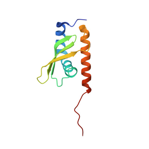
    Heme has a critical role in the chemical framework of the cell as an essential protein cofactor and signaling molecule that controls diverse processes and molecular interactions. Using a phylogenomics-based approach and complementary structural techniques, we identify a family of dimeric hemoproteins comprising a domain of unknown function DUF2470. The heme iron is axially coordinated by two zinc-bound histidine residues, forming a distinct two-fold symmetric zinc-histidine-iron-histidine-zinc site. Together with structure-guided in vitro and in vivo experiments, we further demonstrate the existence of a functional link between heme binding by Dri1 (Domain related to iron 1, formerly ssr1698) and post-translational regulation of succinate dehydrogenase in the cyanobacterium Synechocystis, suggesting an iron-dependent regulatory link between photosynthesis and respiration. Given the ubiquity of proteins containing homologous domains and connections to heme metabolism across eukaryotes and prokaryotes, we propose that DRI (Domain Related to Iron; formerly DUF2470) functions at the molecular level as a heme-dependent regulatory domain.


  • Organizational Affiliation
    • Biology Department, Brookhaven National Laboratory, Upton, NY, USA.

Macromolecule Content 

  • Total Structure Weight: 23.1 kDa 
  • Atom Count: 1,523 
  • Modeled Residue Count: 196 
  • Deposited Residue Count: 206 
  • Unique protein chains: 1

Macromolecules

Find similar proteins by:|  3D Structure
Entity ID: 1
MoleculeChains  Sequence LengthOrganismDetailsImage
Ssr1698 protein
A, B
103Synechocystis sp. PCC 6803Mutation(s): 1 
Gene Names: ssr1698
UniProt
Find proteins for P73129 (Synechocystis sp. (strain ATCC 27184 / PCC 6803 / Kazusa))
Explore P73129 
Go to UniProtKB:  P73129
Entity Groups
Sequence Clusters30% Identity50% Identity70% Identity90% Identity95% Identity100% Identity
UniProt GroupP73129
Sequence Annotations
Expand
Reference Sequence

Small Molecules

Ligands 1 Unique
IDChains Name / Formula / InChI Key2D Diagram3D Interactions
HEB
(Subject of Investigation/LOI)

Query on HEB


Download:Ideal Coordinates CCD File
C [auth A]HEME B/C
C34 H34 Fe N4 O4
NEGHHAJBRZGUAY-RGGAHWMASA-L

Experimental Data & Validation

Experimental Data

  • Method: X-RAY DIFFRACTION
  • Resolution: 2.85 Å
  • R-Value Free:  0.271 (Depositor), 0.263 (DCC) 
  • R-Value Work:  0.219 (Depositor), 0.215 (DCC) 
  • R-Value Observed: 0.222 (Depositor) 
Space Group: P 65 2 2
Unit Cell:
Length ( Å )Angle ( ˚ )
a = 74.659α = 90
b = 74.659β = 90
c = 211.188γ = 120
Software Package:
Software NamePurpose
PHENIXrefinement
autoPROCdata processing
Aimlessdata scaling
XDSdata reduction
PHASERphasing
Cootmodel building

Structure Validation

View Full Validation Report


Ligand Structure Quality Assessment 


Entry History 

& Funding Information

Deposition Data


Funding OrganizationLocationGrant Number
Not funded--

Revision History  (Full details and data files)

  • Version 1.0: 2024-04-24
    Type: Initial release