Download Mendeley
Structures of trehalose-6-phosphate synthase, Tps1, from the fungal pathogen Cryptococcus neoformans : a target for novel antifungals.
Washington, E.J., Zhou, Y., Hsu, A.L., Petrovich, M., Tenor, J.L., Toffaletti, D.L., Guan, Z., Perfect, J.R., Borgnia, M.J., Bartesaghi, A., Brennan, R.G.(2024) bioRxiv
- PubMed: 36993618 Search on PubMedSearch on PubMed Central
- DOI: https://doi.org/10.1101/2023.03.14.530545
- Primary Citation Related Structures:
8FHW - PubMed Abstract:
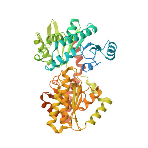
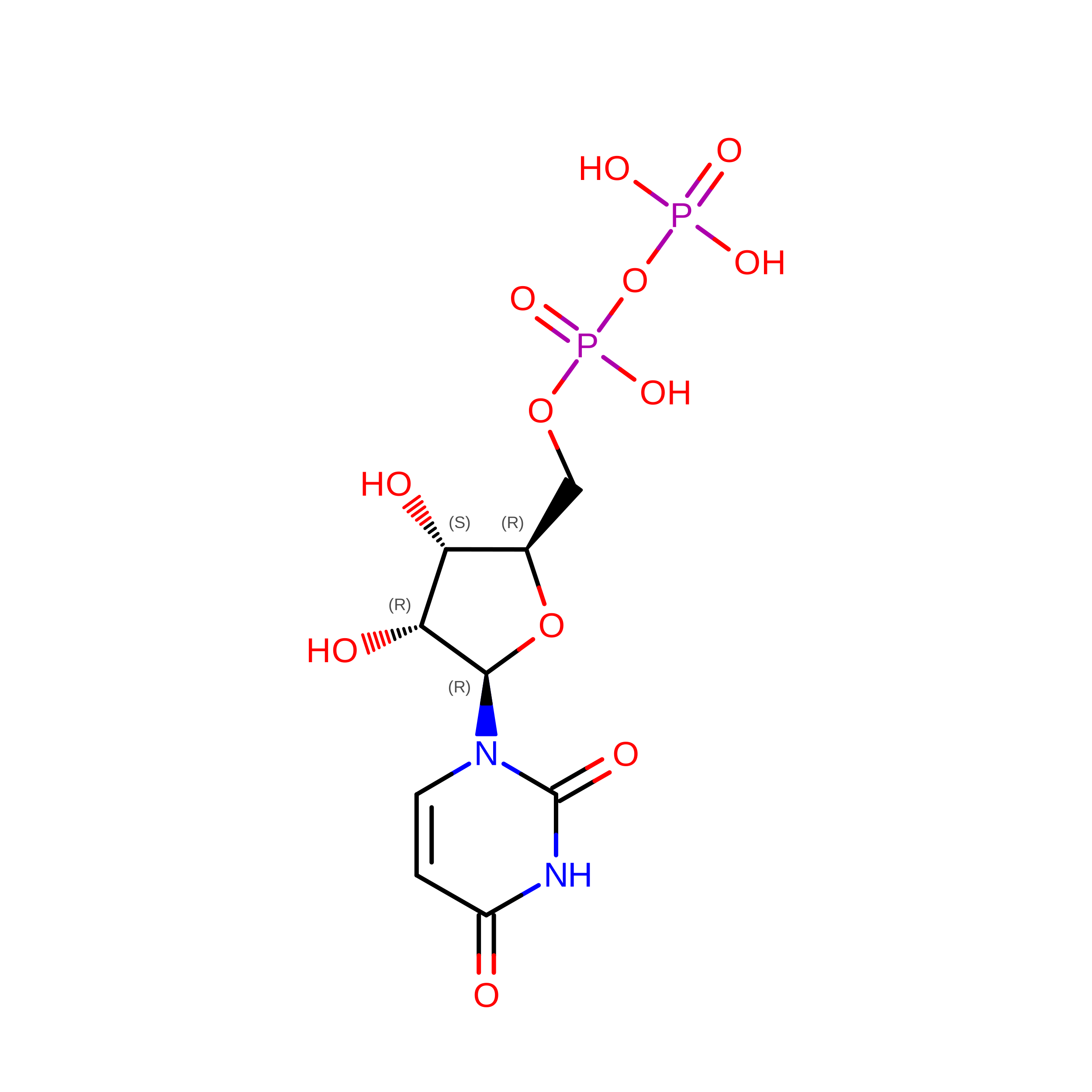
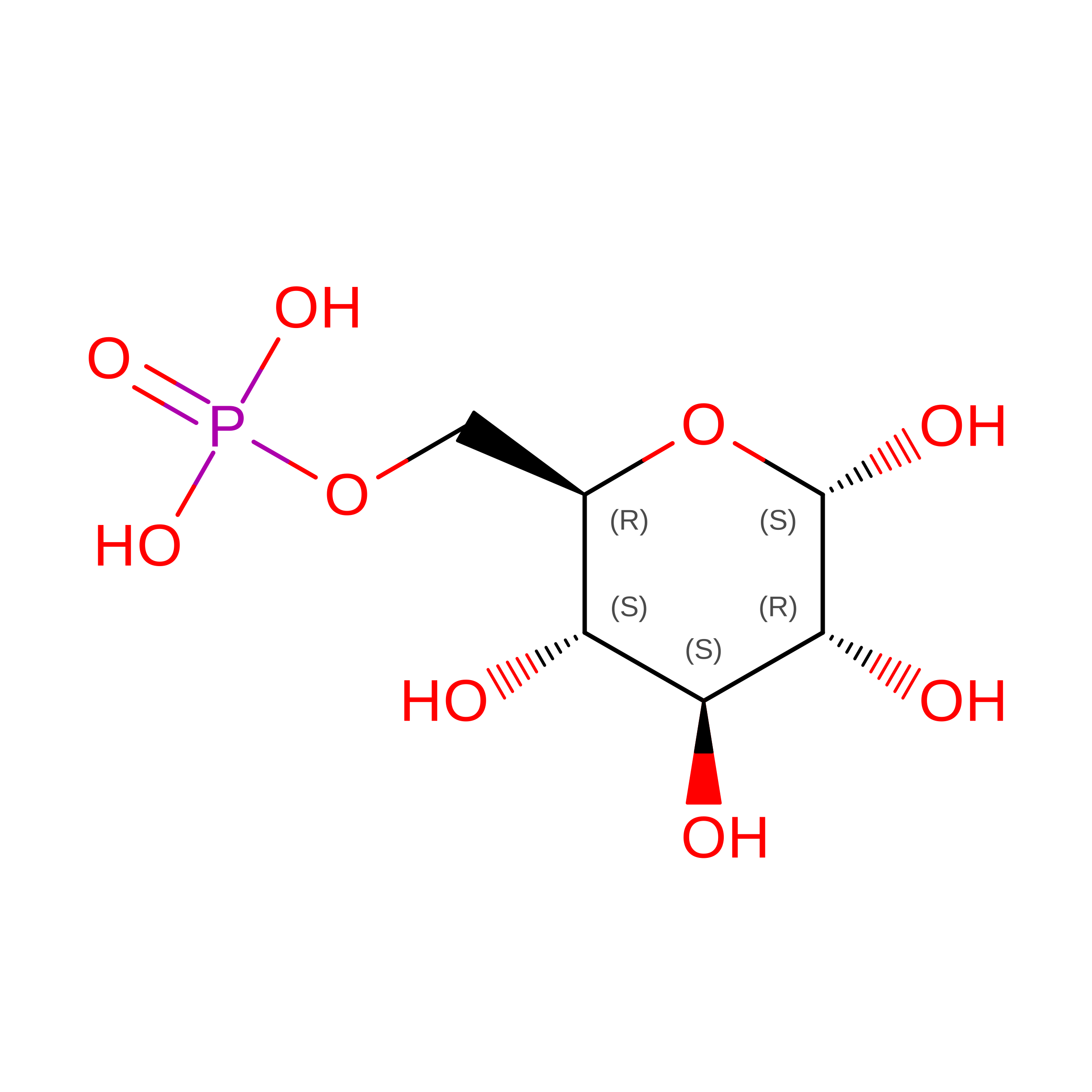
Invasive fungal diseases are a major threat to human health, resulting in more than 1.5 million annual deaths worldwide. The arsenal of antifungal therapeutics remains limited and is in dire need of novel drugs that target additional biosynthetic pathways that are absent from humans. One such pathway involves the biosynthesis of trehalose. Trehalose is a disaccharide that is required for pathogenic fungi to survive in their human hosts. In the first step of trehalose biosynthesis, trehalose-6-phosphate synthase (Tps1) converts UDP-glucose and glucose-6-phosphate to trehalose-6-phosphate. Here, we report the structures of full-length Cryptococcus neoformans Tps1 (CnTps1) in unliganded form and in complex with uridine diphosphate and glucose-6-phosphate. Comparison of these two structures reveals significant movement towards the catalytic pocket by the N-terminus upon ligand binding and identifies residues required for substrate-binding, as well as residues that stabilize the tetramer. Intriguingly, an intrinsically disordered domain (IDD), which is conserved amongst Cryptococcal species and closely related Basidiomycetes, extends from each subunit of the tetramer into the "solvent" but is not visible in density maps. We determined that the IDD is not required for C. neoformans Tps1-dependent thermotolerance and osmotic stress survival. Studies with UDP-galactose highlight the exquisite substrate specificity of CnTps1. In toto , these studies expand our knowledge of trehalose biosynthesis in Cryptococcus and highlight the potential of developing antifungal therapeutics that disrupt the synthesis of this disaccharide or the formation of a functional tetramer and the use of cryo-EM in the structural characterization of CnTps1-ligand/drug complexes. Fungal infections are responsible for over a million deaths worldwide each year. Biosynthesis of a disaccharide, trehalose, is required for multiple pathogenic fungi to transition from the environment to the human host. Enzymes in the trehalose biosynthesis pathway are absent in humans and, therefore, are potentially significant targets for novel antifungal therapeutics. One enzyme in the trehalose biosynthesis is trehalose-6-phosphate synthase (Tps1). Here, we describe the cryo-electron microscopy structures of the CnTps1 homo-tetramer in the unliganded form and in complex with a substrate and a product. These structures and subsequent biochemical analysis reveal key details of substrate-binding residues and substrate specificity. These structures should facilitate structure-guided design of inhibitors against CnTps1.