8FHV

Structure of Lettuce aptamer bound to DFHBI-1T with thallium I ions


Experimental Data Snapshot

  • Method: X-RAY DIFFRACTION
  • Resolution: 2.50 Å
  • R-Value Free: 0.251 
  • R-Value Work: 0.212 
  • R-Value Observed: 0.216 

wwPDB Validation   3D Report Full Report


Ligand Structure Quality Assessment 


This is version 1.1 of the entry. See complete history


Literature

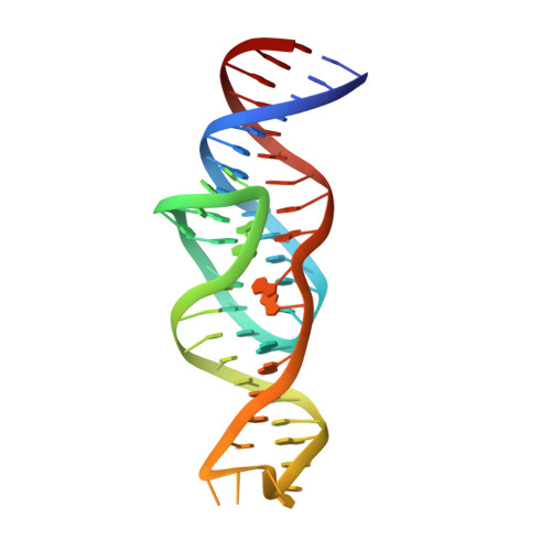
Intricate 3D architecture of a DNA mimic of GFP.

Passalacqua, L.F.M.Banco, M.T.Moon, J.D.Li, X.Jaffrey, S.R.Ferre-D'Amare, A.R.

(2023) Nature 618: 1078-1084

  • DOI: https://doi.org/10.1038/s41586-023-06229-8
  • Primary Citation of Related Structures:  
    8FHV, 8FHX, 8FHZ, 8FI0, 8FI1, 8FI2, 8FI7, 8FI8

  • PubMed Abstract: 

    Numerous studies have shown how RNA molecules can adopt elaborate three-dimensional (3D) architectures 1-3 . By contrast, whether DNA can self-assemble into complex 3D folds capable of sophisticated biochemistry, independent of protein or RNA partners, has remained mysterious. Lettuce is an in vitro-evolved DNA molecule that binds and activates 4 conditional fluorophores derived from GFP. To extend previous structural studies 5,6 of fluorogenic RNAs, GFP and other fluorescent proteins 7 to DNA, we characterize Lettuce-fluorophore complexes by X-ray crystallography and cryogenic electron microscopy. The results reveal that the 53-nucleotide DNA adopts a four-way junction (4WJ) fold. Instead of the canonical L-shaped or H-shaped structures commonly seen 8 in 4WJ RNAs, the four stems of Lettuce form two coaxial stacks that pack co-linearly to form a central G-quadruplex in which the fluorophore binds. This fold is stabilized by stacking, extensive nucleobase hydrogen bonding-including through unusual diagonally stacked bases that bridge successive tiers of the main coaxial stacks of the DNA-and coordination of monovalent and divalent cations. Overall, the structure is more compact than many RNAs of comparable size. Lettuce demonstrates how DNA can form elaborate 3D structures without using RNA-like tertiary interactions and suggests that new principles of nucleic acid organization will be forthcoming from the analysis of complex DNAs.


  • Organizational Affiliation

    Laboratory of Nucleic Acids, National Heart, Lung, and Blood Institute, National Institutes of Health, Bethesda, MD, USA.


Macromolecules
Find similar nucleic acids by:  (by identity cutoff)  |  3D Structure
Entity ID: 1
MoleculeChains LengthOrganismImage
Lettuce DNA aptamer53synthetic construct
Sequence Annotations
Expand
  • Reference Sequence
Small Molecules
Ligands 3 Unique
IDChains Name / Formula / InChI Key2D Diagram3D Interactions
2ZY (Subject of Investigation/LOI)
Query on 2ZY

Download Ideal Coordinates CCD File 
H [auth A](5Z)-5-(3,5-difluoro-4-hydroxybenzylidene)-2-methyl-3-(2,2,2-trifluoroethyl)-3,5-dihydro-4H-imidazol-4-one
C13 H9 F5 N2 O2
AWYCLBWNRONMQC-WMZJFQQLSA-N
TL
Query on TL

Download Ideal Coordinates CCD File 
B [auth A],
C [auth A]
THALLIUM (I) ION
Tl
ZLUSCZLCHQSJRU-UHFFFAOYSA-N
MG
Query on MG

Download Ideal Coordinates CCD File 
D [auth A],
E [auth A],
F [auth A],
G [auth A]
MAGNESIUM ION
Mg
JLVVSXFLKOJNIY-UHFFFAOYSA-N
Experimental Data & Validation

Experimental Data

  • Method: X-RAY DIFFRACTION
  • Resolution: 2.50 Å
  • R-Value Free: 0.251 
  • R-Value Work: 0.212 
  • R-Value Observed: 0.216 
  • Space Group: P 21 21 21
Unit Cell:
Length ( Å )Angle ( ˚ )
a = 24.658α = 90
b = 42.821β = 90
c = 119.531γ = 90
Software Package:
Software NamePurpose
Cootmodel building
PHENIXrefinement
XDSdata reduction
Aimlessdata scaling
AutoSolphasing

Structure Validation

View Full Validation Report


Ligand Structure Quality Assessment 


Entry History & Funding Information

Deposition Data


Funding OrganizationLocationGrant Number
National Institutes of Health/National Heart, Lung, and Blood Institute (NIH/NHLBI)United States--

Revision History  (Full details and data files)

  • Version 1.0: 2023-05-10
    Type: Initial release
  • Version 1.1: 2023-07-05
    Changes: Database references