Selective steroidogenic cytochrome P450 haem iron ligation by steroid-derived isonitriles.
Richard, A.M., Wong, N.R., Harris, K., Sundar, R., Scott, E.E., Pochapsky, T.C.(2023) Commun Chem 6: 183-183
- PubMed: 37660137 Search on PubMedSearch on PubMed Central
- DOI: https://doi.org/10.1038/s42004-023-00994-3
- Primary Citation Related Structures:
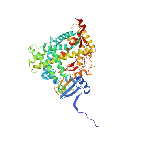
8FDA - PubMed Abstract:
Alkyl isonitriles, R-NC, have previously been shown to ligate the heme (haem) iron of cytochromes P450 in both accessible oxidation states (ferrous, Fe 2+ , and ferric, Fe 3+ ). Herein, the preparation of four steroid-derived isonitriles and their interactions with several P450s, including the steroidogenic CYP17A1 and CYP106A2, as well as the more promiscuous drug metabolizers CYP3A4 and CYP2D6, is described. It was found that successful ligation of the heme iron by the isonitrile functionality for a given P450 depends on both the position and stereochemistry of the isonitrile on the steroid skeleton. Spectral studies indicate that isonitrile ligation of the ferric heme is stable upon reduction to the ferrous form, with reoxidation resulting in the original complex. A crystallographic structure of CYP17A1 with an isonitrile derived from pregnanalone further confirmed the interaction and identified the absolute stereochemistry of the bound species.
- Chemical Biology Program, University of Michigan, Ann Arbor, 48109, MI, USA.
Organizational Affiliation: