8FC7

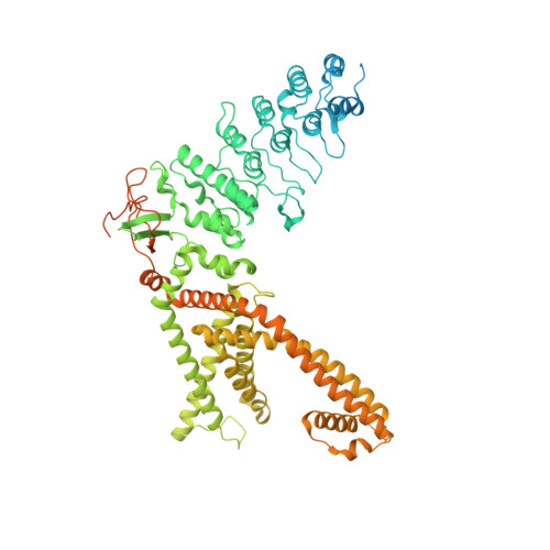
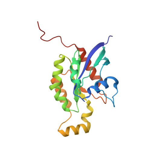
Cryo-EM structure of the human TRPV4 - RhoA in complex with GSK2798745


Experimental Data Snapshot

  • Method: ELECTRON MICROSCOPY
  • Resolution: 3.30 Å
  • Aggregation State: CELL 
  • Reconstruction Method: SINGLE PARTICLE 

wwPDB Validation   3D Report Full Report


This is version 1.0 of the entry. See complete history


Literature

TRPV4-Rho GTPase complex structures reveal mechanisms of gating and disease.

Kwon, D.H.Zhang, F.McCray, B.A.Feng, S.Kumar, M.Sullivan, J.M.Im, W.Sumner, C.J.Lee, S.Y.

(2023) Nat Commun 14: 3732-3732

  • DOI: https://doi.org/10.1038/s41467-023-39345-0
  • Primary Citation of Related Structures:  
    8FC7, 8FC8, 8FC9, 8FCA, 8FCB

  • PubMed Abstract: 

    Crosstalk between ion channels and small GTPases is critical during homeostasis and disease, but little is known about the structural underpinnings of these interactions. TRPV4 is a polymodal, calcium-permeable cation channel that has emerged as a potential therapeutic target in multiple conditions. Gain-of-function mutations also cause hereditary neuromuscular disease. Here, we present cryo-EM structures of human TRPV4 in complex with RhoA in the ligand-free, antagonist-bound closed, and agonist-bound open states. These structures reveal the mechanism of ligand-dependent TRPV4 gating. Channel activation is associated with rigid-body rotation of the intracellular ankyrin repeat domain, but state-dependent interaction with membrane-anchored RhoA constrains this movement. Notably, many residues at the TRPV4-RhoA interface are mutated in disease and perturbing this interface by introducing mutations into either TRPV4 or RhoA increases TRPV4 channel activity. Together, these results suggest that RhoA serves as an auxiliary subunit for TRPV4, regulating TRPV4-mediated calcium homeostasis and disruption of TRPV4-RhoA interactions can lead to TRPV4-related neuromuscular disease. These insights will help facilitate TRPV4 therapeutics development.


  • Organizational Affiliation

    Department of Biochemistry, Duke University School of Medicine, Durham, NC, 27710, USA.


Macromolecules
Find similar proteins by:  (by identity cutoff)  |  3D Structure
Entity ID: 1
MoleculeChains Sequence LengthOrganismDetailsImage
Transient receptor potential cation channel subfamily V member 4A,
F [auth B],
G [auth C],
H [auth D]
901Homo sapiensMutation(s): 0 
Gene Names: TRPV4VRL2VROAC
Membrane Entity: Yes 
UniProt & NIH Common Fund Data Resources
Find proteins for Q9HBA0 (Homo sapiens)
Explore Q9HBA0 
Go to UniProtKB:  Q9HBA0
PHAROS:  Q9HBA0
GTEx:  ENSG00000111199 
Entity Groups  
Sequence Clusters30% Identity50% Identity70% Identity90% Identity95% Identity100% Identity
UniProt GroupQ9HBA0
Sequence Annotations
Expand
  • Reference Sequence
Find similar proteins by:  (by identity cutoff)  |  3D Structure
Entity ID: 2
MoleculeChains Sequence LengthOrganismDetailsImage
Transforming protein RhoAB [auth E],
C [auth F],
D [auth G],
E [auth H]
193Homo sapiensMutation(s): 0 
Gene Names: RHOAARH12ARHARHO12
EC: 3.6.5.2
Membrane Entity: Yes 
UniProt & NIH Common Fund Data Resources
Find proteins for P61586 (Homo sapiens)
Explore P61586 
Go to UniProtKB:  P61586
PHAROS:  P61586
GTEx:  ENSG00000067560 
Entity Groups  
Sequence Clusters30% Identity50% Identity70% Identity90% Identity95% Identity100% Identity
UniProt GroupP61586
Sequence Annotations
Expand
  • Reference Sequence
Small Molecules
Ligands 3 Unique
IDChains Name / Formula / InChI Key2D Diagram3D Interactions
XPW (Subject of Investigation/LOI)
Query on XPW

Download Ideal Coordinates CCD File 
I [auth A],
R [auth B],
S [auth C],
T [auth D]
1-({(5S,7S)-3-[5-(2-hydroxypropan-2-yl)pyrazin-2-yl]-7-methyl-2-oxo-1-oxa-3-azaspiro[4.5]decan-7-yl}methyl)-1H-benzimidazole-6-carbonitrile
C25 H28 N6 O3
URCMKDJBULWNAI-DQEYMECFSA-N
GDP
Query on GDP

Download Ideal Coordinates CCD File 
K [auth E],
M [auth F],
O [auth G],
Q [auth H]
GUANOSINE-5'-DIPHOSPHATE
C10 H15 N5 O11 P2
QGWNDRXFNXRZMB-UUOKFMHZSA-N
MG
Query on MG

Download Ideal Coordinates CCD File 
J [auth E],
L [auth F],
N [auth G],
P [auth H]
MAGNESIUM ION
Mg
JLVVSXFLKOJNIY-UHFFFAOYSA-N
Experimental Data & Validation

Experimental Data

  • Method: ELECTRON MICROSCOPY
  • Resolution: 3.30 Å
  • Aggregation State: CELL 
  • Reconstruction Method: SINGLE PARTICLE 

Structure Validation

View Full Validation Report


Entry History & Funding Information

Deposition Data


Funding OrganizationLocationGrant Number
National Institutes of Health/National Institute of Diabetes and Digestive and Kidney Disease (NIH/NIDDK)United States--

Revision History  (Full details and data files)

  • Version 1.0: 2023-07-12
    Type: Initial release