8FB0

H64Q Myoglobin in complex with acetamide


Experimental Data Snapshot

  • Method: X-RAY DIFFRACTION
  • Resolution: 1.76 Å
  • R-Value Free: 0.194 
  • R-Value Work: 0.163 
  • R-Value Observed: 0.165 

wwPDB Validation   3D Report Full Report


Ligand Structure Quality Assessment 


This is version 1.2 of the entry. See complete history


Literature

Interactions of metronidazole and chloramphenicol with myoglobin: Crystal structure of a Mb-acetamide product.

Powell, S.M.Prather, K.Y.Nguyen, N.Thomas, L.M.Richter-Addo, G.B.

(2023) J Porphyr Phthalocyanines 27: 1142-1147

  • DOI: https://doi.org/10.1142/s1088424623500700
  • Primary Citation of Related Structures:  
    8FB0

  • PubMed Abstract: 

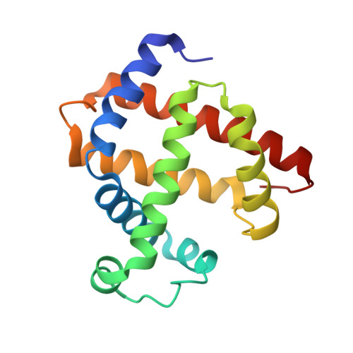
    Nitroorganics present a general concern for a safe environment due to their health hazards. However, some nitroorganics such as metronidazole (Mtz) and chloramphenicol (CAM) also possess medicinal value. Mtz and CAM can undergo reductive bioactivation presumably via their nitroso derivatives. We show, using UV-vis spectroscopy, that sperm whale myoglobin (swMb) and its distal pocket mutants retaining H-bonding capacity react with Mtz in the presence of dithionite to generate products with spectra suggestive of the Fe-bound nitroso (Fe-RNO; λ max ~420 nm) forms. We have crystallized and solved the X-ray crystal structure of an H64Q swMb-acetamide compound to 1.76 Å resolution; formation of this compound results from the serendipitous crystallographic trapping, by the heme center, of acetamide from the reductive decomposition of Mtz. Only one of the swMb proteins, namely H64Q swMb with a relatively flexible Gln64 residue, reacted with CAM presumably due to the bulky nature of CAM that generally may restrict its access to the heme site.


  • Organizational Affiliation

    Price Family Foundation Institute of Structural Biology, Stephenson Life Sciences Research Center, and Department of Chemistry and Biochemistry, University of Oklahoma, 101 Stephenson Parkway, Norman, OK, U.S.A. 73019.


Macromolecules
Find similar proteins by:  (by identity cutoff)  |  3D Structure
Entity ID: 1
MoleculeChains Sequence LengthOrganismDetailsImage
Myoglobin154Physeter catodonMutation(s): 2 
Gene Names: MB
UniProt
Find proteins for P02185 (Physeter macrocephalus)
Explore P02185 
Go to UniProtKB:  P02185
Entity Groups  
Sequence Clusters30% Identity50% Identity70% Identity90% Identity95% Identity100% Identity
UniProt GroupP02185
Sequence Annotations
Expand
  • Reference Sequence
Small Molecules
Ligands 4 Unique
IDChains Name / Formula / InChI Key2D Diagram3D Interactions
HEM
Query on HEM

Download Ideal Coordinates CCD File 
B [auth A]PROTOPORPHYRIN IX CONTAINING FE
C34 H32 Fe N4 O4
KABFMIBPWCXCRK-RGGAHWMASA-L
SO4
Query on SO4

Download Ideal Coordinates CCD File 
F [auth A],
G [auth A],
H [auth A],
I [auth A]
SULFATE ION
O4 S
QAOWNCQODCNURD-UHFFFAOYSA-L
GOL
Query on GOL

Download Ideal Coordinates CCD File 
D [auth A],
E [auth A]
GLYCEROL
C3 H8 O3
PEDCQBHIVMGVHV-UHFFFAOYSA-N
ACM (Subject of Investigation/LOI)
Query on ACM

Download Ideal Coordinates CCD File 
C [auth A]ACETAMIDE
C2 H5 N O
DLFVBJFMPXGRIB-UHFFFAOYSA-N
Experimental Data & Validation

Experimental Data

  • Method: X-RAY DIFFRACTION
  • Resolution: 1.76 Å
  • R-Value Free: 0.194 
  • R-Value Work: 0.163 
  • R-Value Observed: 0.165 
  • Space Group: P 6
Unit Cell:
Length ( Å )Angle ( ˚ )
a = 90.288α = 90
b = 90.288β = 90
c = 45.255γ = 120
Software Package:
Software NamePurpose
PHENIXrefinement
HKL-3000data collection
HKL-3000data reduction
HKL-3000data scaling
PHASERphasing

Structure Validation

View Full Validation Report


Ligand Structure Quality Assessment 


Entry History & Funding Information

Deposition Data


Funding OrganizationLocationGrant Number
National Science Foundation (NSF, United States)United States--

Revision History  (Full details and data files)

  • Version 1.0: 2023-06-21
    Type: Initial release
  • Version 1.1: 2023-10-25
    Changes: Data collection, Refinement description
  • Version 1.2: 2023-11-01
    Changes: Database references