8F69 | pdb_00008f69

Crystal structure of murine PolG2 dimer bound to DNA


Experimental Data Snapshot

  • Method: X-RAY DIFFRACTION
  • Resolution: 2.20 Å
  • R-Value Free: 
    0.228 (Depositor), 0.243 (DCC) 
  • R-Value Work: 
    0.194 (Depositor) 
  • R-Value Observed: 
    0.195 (Depositor) 

Starting Model: experimental
View more details

wwPDB Validation 3D Report Full Report

Validation slider image for 8F69

This is version 1.3 of the entry. See complete history

Literature

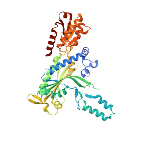
Structure-specific roles for PolG2-DNA complexes in maintenance and replication of mitochondrial DNA.

Wojtaszek, J.L.Hoff, K.E.Longley, M.J.Kaur, P.Andres, S.N.Wang, H.Williams, R.S.Copeland, W.C.

(2023) Nucleic Acids Res 51: 9716-9732

  • DOI: https://doi.org/10.1093/nar/gkad679
  • Primary Citation Related Structures: 
    8F69, 8F6B

  • PubMed Abstract: 

    The homodimeric PolG2 accessory subunit of the mitochondrial DNA polymerase gamma (Pol γ) enhances DNA binding and processive DNA synthesis by the PolG catalytic subunit. PolG2 also directly binds DNA, although the underlying molecular basis and functional significance are unknown. Here, data from Atomic Force Microscopy (AFM) and X-ray structures of PolG2-DNA complexes define dimeric and hexameric PolG2 DNA binding modes. Targeted disruption of PolG2 DNA-binding interfaces impairs processive DNA synthesis without diminishing Pol γ subunit affinities. In addition, a structure-specific DNA-binding role for PolG2 oligomers is supported by X-ray structures and AFM showing that oligomeric PolG2 localizes to DNA crossings and targets forked DNA structures resembling the mitochondrial D-loop. Overall, data indicate that PolG2 DNA binding has both PolG-dependent and -independent functions in mitochondrial DNA replication and maintenance, which provide new insight into molecular defects associated with PolG2 disruption in mitochondrial disease.


  • Organizational Affiliation
    • Genome Integrity and Structural Biology Laboratory, National Institute of Environmental Health Sciences, National Institutes of Health, Research Triangle Park, NC 27709, USA.

Macromolecule Content 

  • Total Structure Weight: 108.8 kDa 
  • Atom Count: 7,352 
  • Modeled Residue Count: 850 
  • Deposited Residue Count: 892 
  • Unique protein chains: 1
  • Unique nucleic acid chains: 1

Macromolecules


Find similar proteins by:|  3D Structure
Entity ID: 1
MoleculeChains  Sequence LengthOrganismDetailsImage
DNA polymerase subunit gamma-2, mitochondrialA [auth B],
B [auth A]
428Mus musculusMutation(s): 0 
Gene Names: Polg2
UniProt & NIH Common Fund Data Resources
Find proteins for Q9QZM2 (Mus musculus)
Explore Q9QZM2 
Go to UniProtKB:  Q9QZM2
IMPC:  MGI:1354947
Entity Groups
Sequence Clusters30% Identity50% Identity70% Identity90% Identity95% Identity100% Identity
UniProt GroupQ9QZM2
Sequence Annotations
Expand
Reference Sequence
Find similar nucleic acids by:  Sequence
Entity ID: 2
MoleculeChains LengthOrganismImage
DNA (5'-D(*CP*TP*GP*GP*TP*AP*GP*GP*CP*GP*CP*CP*TP*AP*CP*CP*AP*G)-3')
C, D
18Homo sapiens
Sequence Annotations
Expand
Reference Sequence

Small Molecules

Ligands 3 Unique
IDChains Name / Formula / InChI Key2D Diagram3D Interactions
MES

Query on MES


Download:Ideal Coordinates CCD File
O [auth A]2-(N-MORPHOLINO)-ETHANESULFONIC ACID
C6 H13 N O4 S
SXGZJKUKBWWHRA-UHFFFAOYSA-N
MPD

Query on MPD


Download:Ideal Coordinates CCD File
F [auth B],
P [auth A]
(4S)-2-METHYL-2,4-PENTANEDIOL
C6 H14 O2
SVTBMSDMJJWYQN-YFKPBYRVSA-N
EDO

Query on EDO


Download:Ideal Coordinates CCD File
E [auth B]
G [auth B]
H [auth B]
I [auth B]
J [auth B]
E [auth B],
G [auth B],
H [auth B],
I [auth B],
J [auth B],
K [auth B],
L [auth B],
M [auth A],
N [auth A],
Q [auth A],
R [auth A],
S [auth A],
T [auth A],
U [auth A]
1,2-ETHANEDIOL
C2 H6 O2
LYCAIKOWRPUZTN-UHFFFAOYSA-N

Experimental Data & Validation

Experimental Data

  • Method: X-RAY DIFFRACTION
  • Resolution: 2.20 Å
  • R-Value Free:  0.228 (Depositor), 0.243 (DCC) 
  • R-Value Work:  0.194 (Depositor) 
  • R-Value Observed: 0.195 (Depositor) 
Space Group: P 41 21 2
Unit Cell:
Length ( Å )Angle ( ˚ )
a = 88.027α = 90
b = 88.027β = 90
c = 343.17γ = 90
Software Package:
Software NamePurpose
PHENIXrefinement
HKL-2000data reduction
HKL-2000data scaling
PHENIXphasing
Cootmodel building

Structure Validation

View Full Validation Report


Entry History 

& Funding Information

Deposition Data


Funding OrganizationLocationGrant Number
National Institutes of Health/National Institute of Environmental Health Sciences (NIH/NIEHS)United States1Z01ES102765

Revision History  (Full details and data files)

  • Version 1.0: 2023-08-09
    Type: Initial release
  • Version 1.1: 2023-08-30
    Changes: Data collection, Database references
  • Version 1.2: 2023-10-25
    Changes: Database references
  • Version 1.3: 2026-03-04
    Changes: Refinement description, Structure summary