Structural and Functional Characterization of Mycobacterium tuberculosis Homoserine Transacetylase.
Sharma, S., Jayasinghe, Y.P., Mishra, N.K., Orimoloye, M.O., Wong, T.Y., Dalluge, J.J., Ronning, D.R., Aldrich, C.C.(2023) ACS Infect Dis 9: 540-553
- PubMed: 36753622 Search on PubMed
- DOI: https://doi.org/10.1021/acsinfecdis.2c00541
- Primary Citation Related Structures:
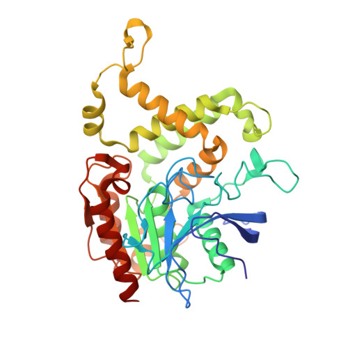
7RYT, 8F2L - PubMed Abstract:
Mycobacterium tuberculosis ( Mtb ) lacking functional homoserine transacetylase (HTA) is compromised in methionine biosynthesis, protein synthesis, and in the activity of multiple essential S -adenosyl-l-methionine-dependent enzymes. Additionally, deficient mutants are further disarmed by the toxic accumulation of lysine due to a redirection of the metabolic flux toward the lysine biosynthetic pathway. Studies with deletion mutants and crystallographic studies of the apoenzyme have, respectively, validated Mtb HTA as an essential enzyme and revealed a ligandable binding site. Seeking a mechanistic characterization of this enzyme, we report crucial structural details and comprehensive functional characterization of Mtb HTA. Crystallographic and mass spectral observation of the acetylated HTA intermediate and initial velocity studies were consistent with a ping-pong kinetic mechanism. Wild-type HTA and its site-directed mutants were kinetically characterized with a panel of natural and alternative substrates to understand substrate specificity and identify critical residues for catalysis. Titration experiments using fluorescence quenching showed that both substrates─acetyl-CoA and l-homoserine─engage in a strong and weak binding interaction with HTA. Additionally, substrate inhibition by acetyl-CoA and product inhibition by CoA and O -acetyl-l-homoserine were proposed to form the basis of a feedback regulation mechanism. By furnishing key mechanistic and structural information, these studies provide a foundation for structure-based design efforts around this attractive Mtb target.
- Department of Medicinal Chemistry, College of Pharmacy, University of Minnesota, Minneapolis, Minnesota 55455, United States.
Organizational Affiliation: