8F0M

Monobody 12D5 bound to KRAS(G12D)


Experimental Data Snapshot

  • Method: X-RAY DIFFRACTION
  • Resolution: 2.44 Å
  • R-Value Free: 0.245 
  • R-Value Work: 0.201 
  • R-Value Observed: 0.203 

wwPDB Validation   3D Report Full Report


Ligand Structure Quality Assessment 


This is version 1.1 of the entry. See complete history


Literature

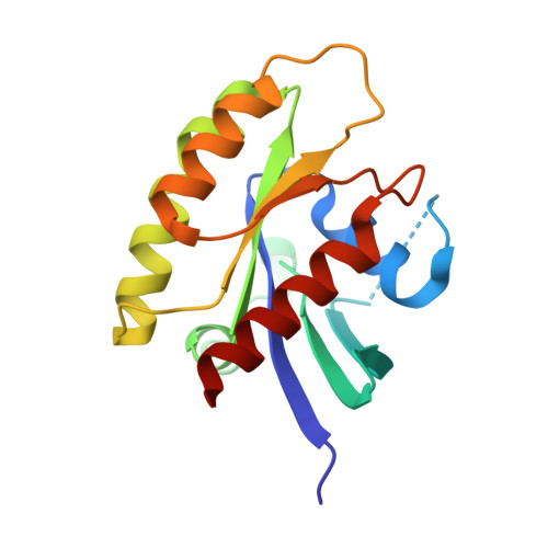
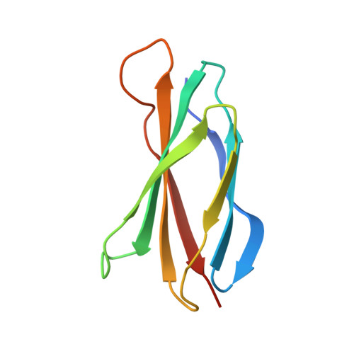
Exploring switch II pocket conformation of KRAS(G12D) with mutant-selective monobody inhibitors.

Akkapeddi, P.Hattori, T.Khan, I.Glasser, E.Koide, A.Ketavarapu, G.Whaby, M.Zuberi, M.Teng, K.W.Lefler, J.Maso, L.Bang, I.Ostrowski, M.C.O'Bryan, J.P.Koide, S.

(2023) Proc Natl Acad Sci U S A 120: e2302485120-e2302485120

  • DOI: https://doi.org/10.1073/pnas.2302485120
  • Primary Citation of Related Structures:  
    8EZG, 8F0M

  • PubMed Abstract: 

    The G12D mutation is among the most common KRAS mutations associated with cancer, in particular, pancreatic cancer. Here, we have developed monobodies, small synthetic binding proteins, that are selective to KRAS(G12D) over KRAS(wild type) and other oncogenic KRAS mutations, as well as over the G12D mutation in HRAS and NRAS. Crystallographic studies revealed that, similar to other KRAS mutant-selective inhibitors, the initial monobody bound to the S-II pocket, the groove between switch II and α3 helix, and captured this pocket in the most widely open form reported to date. Unlike other G12D-selective polypeptides reported to date, the monobody used its backbone NH group to directly recognize the side chain of KRAS Asp12, a feature that closely resembles that of a small-molecule inhibitor, MTRX1133. The monobody also directly interacted with H95, a residue not conserved in RAS isoforms. These features rationalize the high selectivity toward the G12D mutant and the KRAS isoform. Structure-guided affinity maturation resulted in monobodies with low nM K D values. Deep mutational scanning of a monobody generated hundreds of functional and nonfunctional single-point mutants, which identified crucial residues for binding and those that contributed to the selectivity toward the GTP- and GDP-bound states. When expressed in cells as genetically encoded reagents, these monobodies engaged selectively with KRAS(G12D) and inhibited KRAS(G12D)-mediated signaling and tumorigenesis. These results further illustrate the plasticity of the S-II pocket, which may be exploited for the design of next-generation KRAS(G12D)-selective inhibitors.


  • Organizational Affiliation

    Laura and Isaac Perlmutter Cancer Center, New York University Langone Health, New York, NY 10016.


Macromolecules
Find similar proteins by:  (by identity cutoff)  |  3D Structure
Entity ID: 1
MoleculeChains Sequence LengthOrganismDetailsImage
Isoform 2B of GTPase KRas
A, C
171Homo sapiensMutation(s): 4 
Gene Names: KRASKRAS2RASK2
EC: 3.6.5.2
UniProt & NIH Common Fund Data Resources
Find proteins for P01116 (Homo sapiens)
Explore P01116 
Go to UniProtKB:  P01116
PHAROS:  P01116
GTEx:  ENSG00000133703 
Entity Groups  
Sequence Clusters30% Identity50% Identity70% Identity90% Identity95% Identity100% Identity
UniProt GroupP01116
Sequence Annotations
Expand
  • Reference Sequence
Find similar proteins by:  (by identity cutoff)  |  3D Structure
Entity ID: 2
MoleculeChains Sequence LengthOrganismDetailsImage
Monobody 12D5
B, D
97Homo sapiensMutation(s): 0 
Entity Groups  
Sequence Clusters30% Identity50% Identity70% Identity90% Identity95% Identity100% Identity
Sequence Annotations
Expand
  • Reference Sequence
Small Molecules
Ligands 4 Unique
IDChains Name / Formula / InChI Key2D Diagram3D Interactions
GSP (Subject of Investigation/LOI)
Query on GSP

Download Ideal Coordinates CCD File 
J [auth C]5'-GUANOSINE-DIPHOSPHATE-MONOTHIOPHOSPHATE
C10 H16 N5 O13 P3 S
XOFLBQFBSOEHOG-UUOKFMHZSA-N
GDP (Subject of Investigation/LOI)
Query on GDP

Download Ideal Coordinates CCD File 
F [auth A]GUANOSINE-5'-DIPHOSPHATE
C10 H15 N5 O11 P2
QGWNDRXFNXRZMB-UUOKFMHZSA-N
GLC
Query on GLC

Download Ideal Coordinates CCD File 
G [auth A],
H [auth B]
alpha-D-glucopyranose
C6 H12 O6
WQZGKKKJIJFFOK-DVKNGEFBSA-N
MG
Query on MG

Download Ideal Coordinates CCD File 
E [auth A],
I [auth C]
MAGNESIUM ION
Mg
JLVVSXFLKOJNIY-UHFFFAOYSA-N
Experimental Data & Validation

Experimental Data

  • Method: X-RAY DIFFRACTION
  • Resolution: 2.44 Å
  • R-Value Free: 0.245 
  • R-Value Work: 0.201 
  • R-Value Observed: 0.203 
  • Space Group: P 21 2 21
Unit Cell:
Length ( Å )Angle ( ˚ )
a = 151.386α = 90
b = 44.733β = 90
c = 88.162γ = 90
Software Package:
Software NamePurpose
PHENIXrefinement
PDB-REDOrefinement
HKL-2000data reduction
HKL-2000data scaling
PHASERphasing

Structure Validation

View Full Validation Report


Ligand Structure Quality Assessment 


Entry History & Funding Information

Deposition Data


Funding OrganizationLocationGrant Number
National Institutes of Health/National Cancer Institute (NIH/NCI)United StatesCA194864
National Institutes of Health/National Cancer Institute (NIH/NCI)United StatesCA201717
National Institutes of Health/National Cancer Institute (NIH/NCI)United StatesCA212608
National Institutes of Health/National Cancer Institute (NIH/NCI)United StatesCA246457
National Institutes of Health/National Cancer Institute (NIH/NCI)United StatesCA225131
American Cancer SocietyUnited StatesPF-18-180-01-TBE

Revision History  (Full details and data files)

  • Version 1.0: 2023-07-19
    Type: Initial release
  • Version 1.1: 2023-10-25
    Changes: Data collection, Refinement description