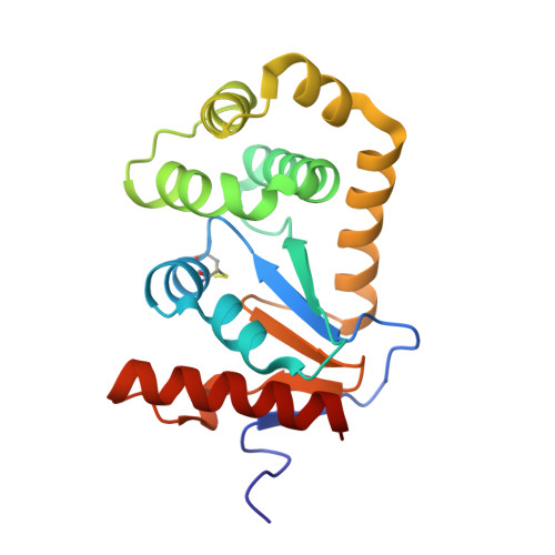
A Buried Water Network Modulates the Activity of the Escherichia coli Disulphide Catalyst DsbA.
Wang, G., Qin, J., Verderosa, A.D., Hor, L., Santos-Martin, C., Paxman, J.J., Martin, J.L., Totsika, M., Heras, B.(2023) Antioxidants (Basel) 12
- PubMed: 36829940
- DOI: https://doi.org/10.3390/antiox12020380
- Primary Citation of Related Structures:
8EOC, 8EQO, 8EQP, 8EQQ, 8EQR - PubMed Abstract:
The formation of disulphide bonds is an essential step in the folding of many proteins that enter the secretory pathway; therefore, it is not surprising that eukaryotic and prokaryotic organisms have dedicated enzymatic systems to catalyse this process. In bacteria, one such enzyme is disulphide bond-forming protein A (DsbA), a thioredoxin-like thiol oxidase that catalyses the oxidative folding of proteins required for virulence and fitness. A large body of work on DsbA proteins, particularly Escherichia coli DsbA (EcDsbA), has demonstrated the key role that the Cys 30 -XX-Cys 33 catalytic motif and its unique redox properties play in the thiol oxidase activity of this enzyme. Using mutational and functional analyses, here we identify that a set of charged residues, which form an acidic groove on the non-catalytic face of the enzyme, further modulate the activity of EcDsbA. Our high-resolution structures indicate that these residues form a water-mediated proton wire that can transfer protons from the bulk solvent to the active site. Our results support the view that proton shuffling may facilitate the stabilisation of the buried Cys 33 thiolate formed during the redox reaction and promote the correct direction of the EcDsbA-substrate thiol-disulphide exchange. Comparison with other proteins of the same class and proteins of the thioredoxin-superfamily in general suggest that a proton relay system appears to be a conserved catalytic feature among this widespread superfamily of proteins. Furthermore, this study also indicates that the acidic groove of DsbA could be a promising allosteric site to develop novel DsbA inhibitors as antibacterial therapeutics.
- Department of Biochemistry and Chemistry, La Trobe Institute for Molecular Science, La Trobe University, Melbourne, VIC 3086, Australia.
Organizational Affiliation:


