8EI3

Crystal structure of VHL in complex with H313, a Helicon Polypeptide


Experimental Data Snapshot

  • Method: X-RAY DIFFRACTION
  • Resolution: 3.49 Å
  • R-Value Free: 0.285 
  • R-Value Work: 0.216 
  • R-Value Observed: 0.219 

wwPDB Validation   3D Report Full Report


Ligand Structure Quality Assessment 


This is version 1.1 of the entry. See complete history


Literature

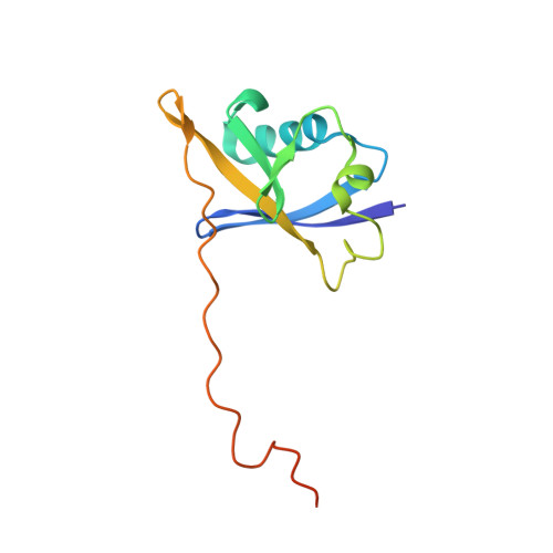
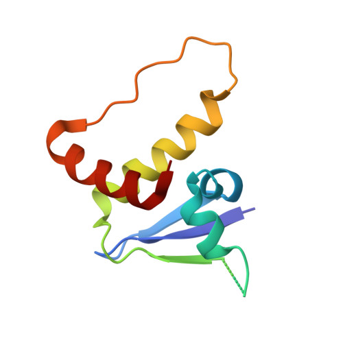
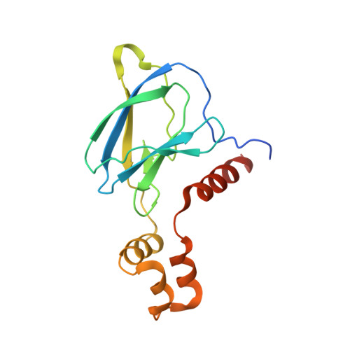
Recognition and reprogramming of E3 ubiquitin ligase surfaces by alpha-helical peptides.

Tokareva, O.S.Li, K.Travaline, T.L.Thomson, T.M.Swiecicki, J.M.Moussa, M.Ramirez, J.D.Litchman, S.Verdine, G.L.McGee, J.H.

(2023) Nat Commun 14: 6992-6992

  • DOI: https://doi.org/10.1038/s41467-023-42395-z
  • Primary Citation of Related Structures:  
    8EHZ, 8EI0, 8EI1, 8EI2, 8EI3, 8EI4, 8EI5, 8EI6, 8EI7, 8EI8, 8EI9, 8EIA, 8EIB, 8EIC

  • PubMed Abstract: 

    Molecules that induce novel interactions between proteins hold great promise for the study of biological systems and the development of therapeutics, but their discovery has been limited by the complexities of rationally designing interactions between three components, and because known binders to each protein are typically required to inform initial designs. Here, we report a general and rapid method for discovering α-helically constrained (Helicon) polypeptides that cooperatively induce the interaction between two target proteins without relying on previously known binders or an intrinsic affinity between the proteins. We show that Helicons are capable of binding every major class of E3 ubiquitin ligases, which are of great biological and therapeutic interest but remain largely intractable to targeting by small molecules. We then describe a phage-based screening method for discovering "trimerizer" Helicons, and apply it to reprogram E3s to cooperatively bind an enzyme (PPIA), a transcription factor (TEAD4), and a transcriptional coactivator (β-catenin).


  • Organizational Affiliation

    FOG Pharmaceuticals Inc., Cambridge, MA, USA.


Macromolecules
Find similar proteins by:  (by identity cutoff)  |  3D Structure
Entity ID: 1
MoleculeChains Sequence LengthOrganismDetailsImage
Elongin-B
A, D
118Homo sapiensMutation(s): 0 
Gene Names: ELOBTCEB2
UniProt & NIH Common Fund Data Resources
Find proteins for Q15370 (Homo sapiens)
Explore Q15370 
Go to UniProtKB:  Q15370
PHAROS:  Q15370
GTEx:  ENSG00000103363 
Entity Groups  
Sequence Clusters30% Identity50% Identity70% Identity90% Identity95% Identity100% Identity
UniProt GroupQ15370
Sequence Annotations
Expand
  • Reference Sequence
Find similar proteins by:  (by identity cutoff)  |  3D Structure
Entity ID: 2
MoleculeChains Sequence LengthOrganismDetailsImage
Elongin-C
B, E
96Homo sapiensMutation(s): 0 
Gene Names: ELOCTCEB1
UniProt & NIH Common Fund Data Resources
Find proteins for Q15369 (Homo sapiens)
Explore Q15369 
Go to UniProtKB:  Q15369
PHAROS:  Q15369
GTEx:  ENSG00000154582 
Entity Groups  
Sequence Clusters30% Identity50% Identity70% Identity90% Identity95% Identity100% Identity
UniProt GroupQ15369
Sequence Annotations
Expand
  • Reference Sequence
Find similar proteins by:  (by identity cutoff)  |  3D Structure
Entity ID: 3
MoleculeChains Sequence LengthOrganismDetailsImage
von Hippel-Lindau disease tumor suppressor
C, F
163Homo sapiensMutation(s): 0 
Gene Names: VHL
UniProt & NIH Common Fund Data Resources
Find proteins for P40337 (Homo sapiens)
Explore P40337 
Go to UniProtKB:  P40337
PHAROS:  P40337
GTEx:  ENSG00000134086 
Entity Groups  
Sequence Clusters30% Identity50% Identity70% Identity90% Identity95% Identity100% Identity
UniProt GroupP40337
Sequence Annotations
Expand
  • Reference Sequence

Find similar proteins by:  Sequence   |   3D Structure  

Entity ID: 4
MoleculeChains Sequence LengthOrganismDetailsImage
H31319synthetic constructMutation(s): 0 
Entity Groups  
Sequence Clusters30% Identity50% Identity70% Identity90% Identity95% Identity100% Identity
Sequence Annotations
Expand
  • Reference Sequence
Small Molecules
Ligands 1 Unique
IDChains Name / Formula / InChI Key2D Diagram3D Interactions
WHL
Query on WHL

Download Ideal Coordinates CCD File 
H [auth G]N,N'-(1,4-phenylene)diacetamide
C10 H12 N2 O2
KVEDKKLZCJBVNP-UHFFFAOYSA-N
Experimental Data & Validation

Experimental Data

  • Method: X-RAY DIFFRACTION
  • Resolution: 3.49 Å
  • R-Value Free: 0.285 
  • R-Value Work: 0.216 
  • R-Value Observed: 0.219 
  • Space Group: P 41
Unit Cell:
Length ( Å )Angle ( ˚ )
a = 47.08α = 90
b = 47.08β = 90
c = 359.58γ = 90
Software Package:
Software NamePurpose
PHENIXrefinement
PDB_EXTRACTdata extraction
XDSdata reduction
Aimlessdata scaling
PHASERphasing

Structure Validation

View Full Validation Report


Ligand Structure Quality Assessment 


Entry History & Funding Information

Deposition Data


Funding OrganizationLocationGrant Number
Other privateUnited States--

Revision History  (Full details and data files)

  • Version 1.0: 2023-10-25
    Type: Initial release
  • Version 1.1: 2023-11-15
    Changes: Database references