8EHO | pdb_00008eho

PRRSV-1 PLP2 domain bound to ubiquitin


Experimental Data Snapshot

  • Method: X-RAY DIFFRACTION
  • Resolution: 2.85 Å
  • R-Value Free: 
    0.247 (Depositor), 0.261 (DCC) 
  • R-Value Work: 
    0.212 (Depositor), 0.220 (DCC) 
  • R-Value Observed: 
    0.214 (Depositor) 

Starting Model: experimental
View more details

wwPDB Validation 3D Report Full Report

Validation slider image for 8EHO

This is version 1.2 of the entry. See complete history

Literature

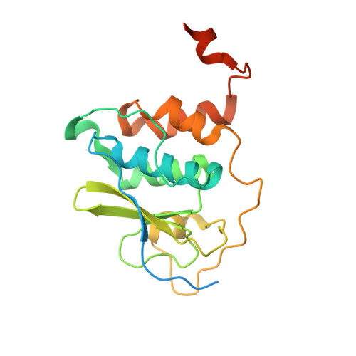
Demonstrating the importance of porcine reproductive and respiratory syndrome virus papain-like protease 2 deubiquitinating activity in viral replication by structure-guided mutagenesis.

Bailey-Elkin, B.A.Knaap, R.C.M.De Silva, A.Boekhoud, I.M.Mous, S.van Vught, N.Khajehpour, M.van den Born, E.Kikkert, M.Mark, B.L.

(2023) PLoS Pathog 19: e1011872-e1011872

  • DOI: https://doi.org/10.1371/journal.ppat.1011872
  • Primary Citation Related Structures: 
    8EHN, 8EHO

  • PubMed Abstract: 

    Deubiquitination of cellular substrates by viral proteases is a mechanism used to interfere with host cellular signaling processes, shared between members of the coronavirus- and arterivirus families. In the case of Arteriviruses, deubiquitinating and polyprotein processing activities are accomplished by the virus-encoded papain-like protease 2 (PLP2). Several studies have implicated the deubiquitinating activity of the porcine reproductive and respiratory syndrome virus (PRRSV) PLP2 in the downregulation of cellular interferon production, however to date, the only arterivirus PLP2 structure described is that of equine arteritis virus (EAV), a distantly related virus. Here we describe the first crystal structure of the PRRSV PLP2 domain both in the presence and absence of its ubiquitin substrate, which reveals unique structural differences in this viral domain compared to PLP2 from EAV. To probe the role of PRRSV PLP2 deubiquitinating activity in host immune evasion, we selectively removed this activity from the domain by mutagenesis and found that the viral domain could no longer downregulate cellular interferon production. Interestingly, unlike EAV, and also unlike the situation for MERS-CoV, we found that recombinant PRRSV carrying PLP2 DUB-specific mutations faces significant selective pressure to revert to wild-type virus in MARC-145 cells, suggesting that the PLP2 DUB activity, which in PRRSV is present as three different versions of viral protein nsp2 expressed during infection, is critically important for PRRSV replication.


  • Organizational Affiliation
    • Department of Microbiology, University of Manitoba, Winnipeg, Manitoba, Canada.

Macromolecule Content 

  • Total Structure Weight: 92.84 kDa 
  • Atom Count: 5,810 
  • Modeled Residue Count: 721 
  • Deposited Residue Count: 837 
  • Unique protein chains: 2

Macromolecules

Find similar proteins by:|  3D Structure
Entity ID: 1
MoleculeChains  Sequence LengthOrganismDetailsImage
Papain-like protease 2
A, C, E
204Porcine reproductive and respiratory syndrome virusMutation(s): 0 
UniProt
Find proteins for W0NX70 (Porcine reproductive and respiratory syndrome virus)
Explore W0NX70 
Go to UniProtKB:  W0NX70
Entity Groups
Sequence Clusters30% Identity50% Identity70% Identity90% Identity95% Identity100% Identity
UniProt GroupW0NX70
Sequence Annotations
Expand
Reference Sequence
Find similar proteins by:|  3D Structure
Entity ID: 2
MoleculeChains  Sequence LengthOrganismDetailsImage
Ubiquitin
B, D, F
75Homo sapiensMutation(s): 0 
Gene Names: UBB
UniProt & NIH Common Fund Data Resources
Find proteins for P0CG47 (Homo sapiens)
Explore P0CG47 
Go to UniProtKB:  P0CG47
PHAROS:  P0CG47
GTEx:  ENSG00000170315 
Entity Groups
Sequence Clusters30% Identity50% Identity70% Identity90% Identity95% Identity100% Identity
UniProt GroupP0CG47
Sequence Annotations
Expand
Reference Sequence

Small Molecules

Ligands 4 Unique
IDChains Name / Formula / InChI Key2D Diagram3D Interactions
GOL

Query on GOL


Download:Ideal Coordinates CCD File
H [auth A]
I [auth A]
M [auth C]
N [auth C]
P [auth D]
H [auth A],
I [auth A],
M [auth C],
N [auth C],
P [auth D],
R [auth E],
S [auth E]
GLYCEROL
C3 H8 O3
PEDCQBHIVMGVHV-UHFFFAOYSA-N
ZN

Query on ZN


Download:Ideal Coordinates CCD File
G [auth A],
L [auth C],
Q [auth E]
ZINC ION
Zn
PTFCDOFLOPIGGS-UHFFFAOYSA-N
NO3

Query on NO3


Download:Ideal Coordinates CCD File
K [auth B]NITRATE ION
N O3
NHNBFGGVMKEFGY-UHFFFAOYSA-N
3CN

Query on 3CN


Download:Ideal Coordinates CCD File
J [auth B],
O [auth D],
T [auth F]
3-AMINOPROPANE
C3 H9 N
WGYKZJWCGVVSQN-UHFFFAOYSA-N

Experimental Data & Validation

Experimental Data

  • Method: X-RAY DIFFRACTION
  • Resolution: 2.85 Å
  • R-Value Free:  0.247 (Depositor), 0.261 (DCC) 
  • R-Value Work:  0.212 (Depositor), 0.220 (DCC) 
  • R-Value Observed: 0.214 (Depositor) 
Space Group: C 2 2 21
Unit Cell:
Length ( Å )Angle ( ˚ )
a = 91.686α = 90
b = 159.492β = 90
c = 147.325γ = 90
Software Package:
Software NamePurpose
PDB-REDOrefinement
XDSdata reduction
XDSdata scaling
PHASERphasing

Structure Validation

View Full Validation Report


Entry History 

& Funding Information

Deposition Data


Funding OrganizationLocationGrant Number
Natural Sciences and Engineering Research Council (NSERC, Canada)CanadaRGPAS-2020-00012
Natural Sciences and Engineering Research Council (NSERC, Canada)CanadaRGPIN-2020-05682

Revision History  (Full details and data files)

  • Version 1.0: 2023-12-06
    Type: Initial release
  • Version 1.1: 2024-01-03
    Changes: Database references
  • Version 1.2: 2026-03-04
    Changes: Derived calculations, Refinement description, Structure summary