8EFR

CryoEM of the soluble OPA1 interfaces with GDP-AlFx bound from the helical assembly on a lipid membrane


Experimental Data Snapshot

  • Method: ELECTRON MICROSCOPY
  • Resolution: 5.48 Å
  • Aggregation State: HELICAL ARRAY 
  • Reconstruction Method: HELICAL 

wwPDB Validation   3D Report Full Report


This is version 1.2 of the entry. See complete history


Literature

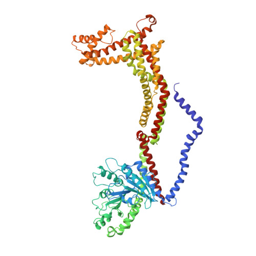
OPA1 helical structures give perspective to mitochondrial dysfunction.

Nyenhuis, S.B.Wu, X.Strub, M.P.Yim, Y.I.Stanton, A.E.Baena, V.Syed, Z.A.Canagarajah, B.Hammer, J.A.Hinshaw, J.E.

(2023) Nature 620: 1109-1116

  • DOI: https://doi.org/10.1038/s41586-023-06462-1
  • Primary Citation of Related Structures:  
    8EEW, 8EF7, 8EFF, 8EFR, 8EFS, 8EFT

  • PubMed Abstract: 

    Dominant optic atrophy is one of the leading causes of childhood blindness. Around 60-80% of cases 1 are caused by mutations of the gene that encodes optic atrophy protein 1 (OPA1), a protein that has a key role in inner mitochondrial membrane fusion and remodelling of cristae and is crucial for the dynamic organization and regulation of mitochondria 2 . Mutations in OPA1 result in the dysregulation of the GTPase-mediated fusion process of the mitochondrial inner and outer membranes 3 . Here we used cryo-electron microscopy methods to solve helical structures of OPA1 assembled on lipid membrane tubes, in the presence and absence of nucleotide. These helical assemblies organize into densely packed protein rungs with minimal inter-rung connectivity, and exhibit nucleotide-dependent dimerization of the GTPase domains-a hallmark of the dynamin superfamily of proteins 4 . OPA1 also contains several unique secondary structures in the paddle domain that strengthen its membrane association, including membrane-inserting helices. The structural features identified in this study shed light on the effects of pathogenic point mutations on protein folding, inter-protein assembly and membrane interactions. Furthermore, mutations that disrupt the assembly interfaces and membrane binding of OPA1 cause mitochondrial fragmentation in cell-based assays, providing evidence of the biological relevance of these interactions.


  • Organizational Affiliation

    Laboratory of Cell and Molecular Biology, National Institute of Diabetes and Digestive and Kidney Diseases, NIH, Bethesda, MD, USA.


Macromolecules
Find similar proteins by:  (by identity cutoff)  |  3D Structure
Entity ID: 1
MoleculeChains Sequence LengthOrganismDetailsImage
Dynamin-like 120 kDa protein, form S1766Homo sapiensMutation(s): 0 
Gene Names: OPA1KIAA0567
UniProt & NIH Common Fund Data Resources
Find proteins for O60313 (Homo sapiens)
Explore O60313 
Go to UniProtKB:  O60313
PHAROS:  O60313
GTEx:  ENSG00000198836 
Entity Groups  
Sequence Clusters30% Identity50% Identity70% Identity90% Identity95% Identity100% Identity
UniProt GroupO60313
Sequence Annotations
Expand
  • Reference Sequence
Small Molecules
Ligands 4 Unique
IDChains Name / Formula / InChI Key2D Diagram3D Interactions
GDP (Subject of Investigation/LOI)
Query on GDP

Download Ideal Coordinates CCD File 
AC [auth Q]
CB [auth N]
DA [auth K]
EC [auth J]
GB [auth G]
AC [auth Q],
CB [auth N],
DA [auth K],
EC [auth J],
GB [auth G],
IA [auth D],
IC [auth R],
KB [auth O],
LA [auth L],
OB [auth H],
PA [auth E],
S [auth A],
SB [auth P],
TA [auth M],
V [auth B],
WB [auth I],
XA [auth F],
Z [auth C]
GUANOSINE-5'-DIPHOSPHATE
C10 H15 N5 O11 P2
QGWNDRXFNXRZMB-UUOKFMHZSA-N
ALF (Subject of Investigation/LOI)
Query on ALF

Download Ideal Coordinates CCD File 
AA [auth C]
BC [auth Q]
DB [auth N]
EA [auth K]
FC [auth J]
AA [auth C],
BC [auth Q],
DB [auth N],
EA [auth K],
FC [auth J],
HB [auth G],
JA [auth D],
JC [auth R],
LB [auth O],
MA [auth L],
PB [auth H],
QA [auth E],
T [auth A],
TB [auth P],
UA [auth M],
W [auth B],
XB [auth I],
YA [auth F]
TETRAFLUOROALUMINATE ION
Al F4
UYOMQIYKOOHAMK-UHFFFAOYSA-J
K (Subject of Investigation/LOI)
Query on K

Download Ideal Coordinates CCD File 
AB [auth F]
BB [auth N]
CA [auth C]
DC [auth Q]
FB [auth N]
AB [auth F],
BB [auth N],
CA [auth C],
DC [auth Q],
FB [auth N],
GA [auth K],
HA [auth D],
HC [auth J],
JB [auth G],
LC [auth R],
NB [auth O],
OA [auth L],
RB [auth H],
SA [auth E],
VB [auth P],
WA [auth M],
Y [auth B],
ZB [auth I]
POTASSIUM ION
K
NPYPAHLBTDXSSS-UHFFFAOYSA-N
MG (Subject of Investigation/LOI)
Query on MG

Download Ideal Coordinates CCD File 
BA [auth C]
CC [auth Q]
EB [auth N]
FA [auth K]
GC [auth J]
BA [auth C],
CC [auth Q],
EB [auth N],
FA [auth K],
GC [auth J],
IB [auth G],
KA [auth D],
KC [auth R],
MB [auth O],
NA [auth L],
QB [auth H],
RA [auth E],
U [auth A],
UB [auth P],
VA [auth M],
X [auth B],
YB [auth I],
ZA [auth F]
MAGNESIUM ION
Mg
JLVVSXFLKOJNIY-UHFFFAOYSA-N
Experimental Data & Validation

Experimental Data

  • Method: ELECTRON MICROSCOPY
  • Resolution: 5.48 Å
  • Aggregation State: HELICAL ARRAY 
  • Reconstruction Method: HELICAL 
EM Software:
TaskSoftware PackageVersion
MODEL REFINEMENTRosetta

Structure Validation

View Full Validation Report


Entry History & Funding Information

Deposition Data


Funding OrganizationLocationGrant Number
National Institutes of Health/National Institute of Diabetes and Digestive and Kidney Disease (NIH/NIDDK)United States--

Revision History  (Full details and data files)

  • Version 1.0: 2023-06-28
    Type: Initial release
  • Version 1.1: 2023-09-06
    Changes: Data collection, Database references
  • Version 1.2: 2023-09-13
    Changes: Database references